Skip to content
  • Our Work
    • Fields
      • Cardiology
      • ENT
      • Gastro
      • Orthopedics
      • Ophthalmology
      • Pulmonology
      • Surgical
      • Urology
      • Other
    • Modalities
      • Endoscopy
      • Medical Segmentation
      • Microscopy
      • Ultrasound
  • Success Stories
  • Insights
    • Magazine
    • Upcoming Events
    • Webinars
    • Meetups
    • News
    • Blog
  • The company
    • About us
    • Careers
Menu
  • Our Work
    • Fields
      • Cardiology
      • ENT
      • Gastro
      • Orthopedics
      • Ophthalmology
      • Pulmonology
      • Surgical
      • Urology
      • Other
    • Modalities
      • Endoscopy
      • Medical Segmentation
      • Microscopy
      • Ultrasound
  • Success Stories
  • Insights
    • Magazine
    • Upcoming Events
    • Webinars
    • Meetups
    • News
    • Blog
  • The company
    • About us
    • Careers
Contact

Tag: Ophthalmology

Zoom-in-Net

Deep Learning in Ophthalmology

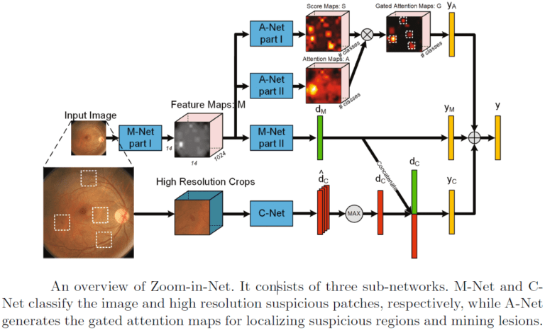
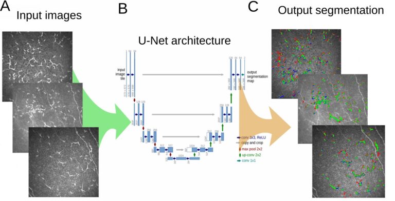
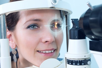
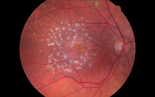
Recent works suggest novel deep learning tools for detection, segmentation and characterization of eye disorders. Accurate segmentation of retinal fundus lesions and anomalies in imaging

Read More
Classification and Segmentation of Dendritic cells

Classification and Segmentation of Dendritic Cells

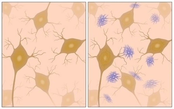
Dry eye disease (DED) is one of the most common ophthalmic disorders. Inflammation of the ocular surface is controlled by corneal antigen-presenting cells called dendritic

Read More
Biometric Detection

Biometric Detection and Measurement

Eyeglasses have been with us since the 13th century, although mass production and affordability to commoner (aside from clerics, scholars and wealthy people) has begun

Read More
Alzheimer's Disease - AD

Degenerative Diseases Detection in the Eye

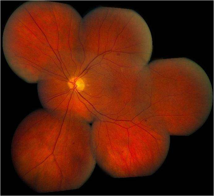
Modern imaging knows how to capture an image to see through the eye tissue transparency and inspect retina, vasculature and neural tissue: this phenomenon is

Read More
Temporary pediatric strabismus in newborn baby

Image processing for pediatric strabismus

Strabismus is a disorder in which the eyes are not properly aligned and point to different directions. When this happens, the “straighter” eye becomes more dominant and the

Read More
ROP - Vessel tortuosity in Retinopathy of Prematurity

ROP: Retinopathy of Prematurity

Retinopathy of prematurity (ROP) is a leading cause of blindness in infants. ROP (or Terry syndrome) is a disease of the eye affecting prematurely-born, low

Read More
Eyelid Drooping - MRD1 and MRD2

Eyelid Drooping – Blepharoptosis

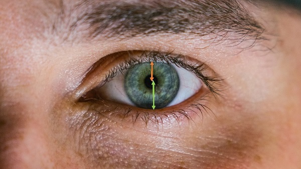
Blepharoptosis, also known as ptosis, is a drooping of the upper eyelid causing a narrowing of the palpebral fissure (or palpebral aperture), which is the

Read More
Fundus image

Retinal images enhancement

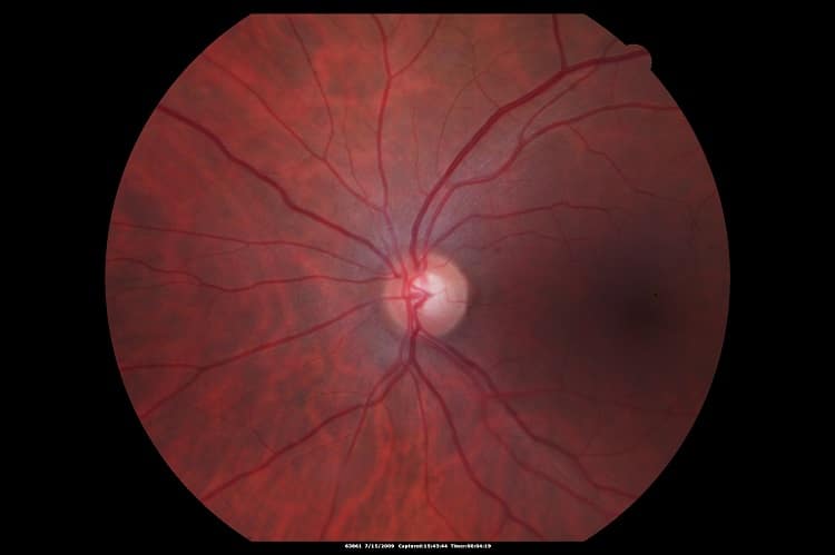
Image enhancement of retinal structures has the potential to facilitate diagnosis of several eye diseases. Retinal disease diagnosis and monitoring often requires very delicate analysis

Read More
Pattern recognition lens markings

Pattern Recognition for Lens Markings Detection

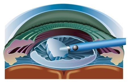
Lens marking refers to the placement of temporary and/or permanent marking, semi-visible laser engraving for the use of lens identification and trademarks, and accurate placement

Read More
Tear film formation and meibomian glands

Meibomian gland dysfunction detection

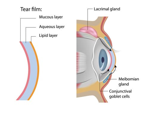
Meibomian gland dysfunction is often seen as an early stage of dry eye syndrome. Indeed, Meibomian glands play a significant role in tears production by

Read More
Eye with Glaucoma

Glaucoma Detection

Glaucoma, a high intraocular pressure (IOP) pathology, leading to damage of the optic nerve, can be better detected using deep learning techniques. When it detects the optical disc (the visible section of the optic nerve), the deep learning algorithm helps assess glaucoma in an automated way, starting from the region of interest and providing a reliable probability for the disease, which the physician will use to support both diagnosis and treatment decisions.

Read More
Giant Retinal Tear

Retinal Detachment Detection

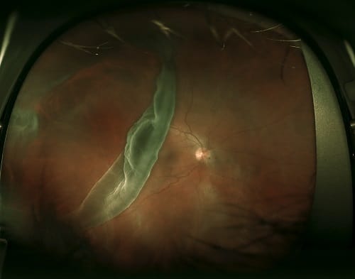
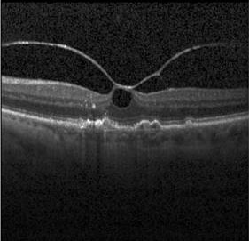
Retinal detachment occurs when part of the retina detaches itself from the pigmented cell layer of the RPE, depriving itself of blood and nutrition: this

Read More
Spectral Domain Optical Coherence Tomography (SD-OCT)

Retinal inner layers segmentation

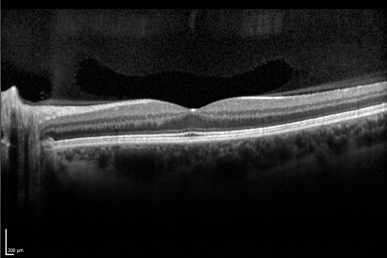
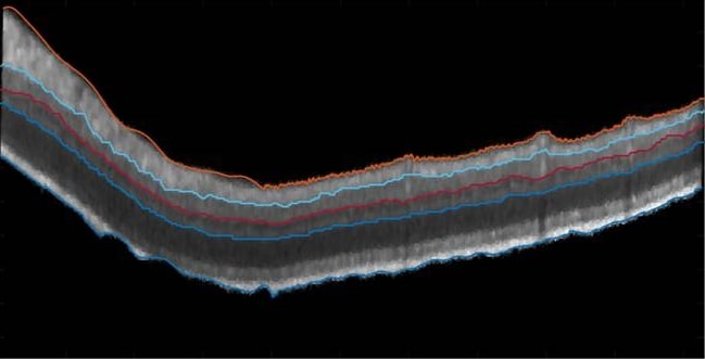
OCT is the only method that can perform noninvasive imaging with non-ionizing radiation and offering relatively good resolution. That is why it has become a

Read More

Diabetic retinopathy screening and microaneurysm detection

Diabetic Retinopathy (DR) is a leading cause of blindness, especially among adults and even more among the elderly segments of the population. It is associated

Read More
Retinal image with smartphone

Retinal images taken with mobile cameras

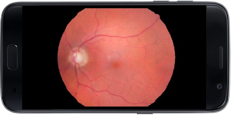
Portable cameras able to help ophthalmologists have been a desired solution for a long time. Among the reasons for the need of an additional device

Read More
SD-OCT image of Geographic Atrophy

Geographic Atrophy Segmentation Using SD-OCT

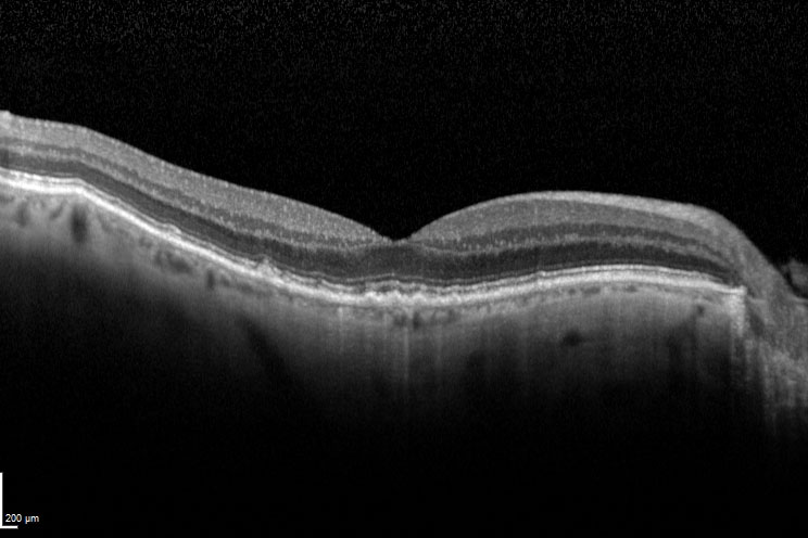
In a previous article, we talked about Geographic Atrophy segmentation in 2D images. This article focuses on how OCT images shed light on the development

Read More
Geographic Atrophy with Drusen

Geographic Atrophy (GA)

Geographic Atrophy (generally called GA) is a case of advanced Dry AMD (Age-related Macular Degeneration) which might lead to vision loss. As a consequence of

Read More
Image Stitching of the Retina

Retina Montage Software

RSIP Vision has developed software which finds common points within the images and reorients them to ‘stitch’ together an accurate panoramic presentation of the retina. Our image stitching technology reconstructs the vascular tree of the retina also when images are acquired at different angles. Its end result enables the ophthalmologist to analyze the patient’s retinal vascular tree through an efficient and non-invasive process.

Read More

Cataract surgery software

Our technology can guide surgeons performing a cataract surgery procedure, by tracking points of interest and their movement. In this way, cuts will be minimized and all operations will be performed with the highest precision. RSIP Vision is able to do that thanks to sophisticated detection and extraction algorithms in the field of image processing, providing the most accurate cataract surgery tool.

Read More
Optical coherence tomography angiography

Optical Coherence Tomography Angiography

Optical coherence tomography angiography uses OCT to produce high-resolution images of the vascular tree in the eye. A fast and non-invasive procedure, it generates high resolution tridimensional images. Stitching multiple images together and increasing image information and clarity, RSIP Vision’s solution allows physicians to quickly and accurately detect pathologies related to vascularization without side effects or clinical hazards.

Read More
Pupil Distance Measurement

Automatic Pupil Distance Measurement

Modern eye glasses need high precision measurements to ensure the best fit. Interpupillary distance, the distance between the projections of the pupil on the cornea, is one of the most important measurements. Sophisticated algorithms, developed by RSIP Vision to determine with high precision the center of vision corresponding to the pupil, have been integrated into machinery used in eye clinics and embedded as an application in portable devices, such as cell phones.

Read More
Multi-Modal Image Registration

Multi-modal registration of retina images

Is it possible to perform combination of fundus images coming from different imaging equipment or technologies and taken from different angles? RSIP Vision apparently can, since we used our expertise in computer vision for ophthalmology to provide software to a client who wanted to combine direct images of the fundus with fluorescein images, which need to be reconstructed before being connected together to form a more detailed image. The end result provides ophthalmologists with detailed images of the retina, ensuring more efficient and accurate patient diagnosis.

Read More
RSIP Vision at American Academy of Ophthalmology (AAO)

Image processing for ophthalmology at AAO 2016

We were glad to participate in the American Academy of Ophthalmology (AAO) annual meeting that took place at the McCormick Place, Chicago, the largest exhibition

Read More
Fluorescein Angiogram

Stabilization in fluorescein angiography

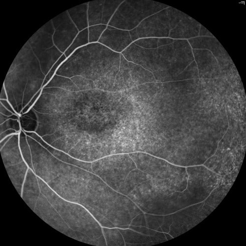
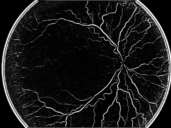
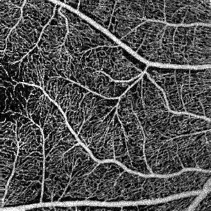
One of the most common tests in ophthalmology is fluorescein angiography: fluorescein is injected in the blood and it moves immediately through the blood vessels

Read More
Diabetic Retinal Screening

Automatic Lesion Detection in Fundus Images

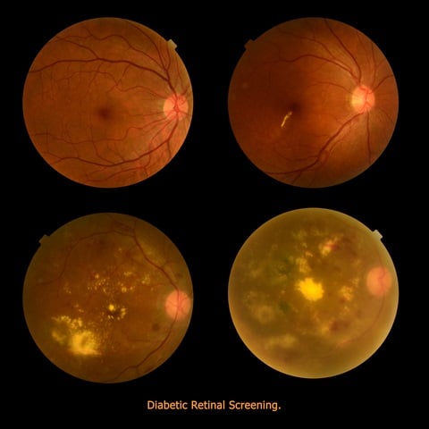
Diabetic Retinopathy (DR) is an eye disease resulting from long-term diabetic condition. About 80% of long-term diabetic patients suffer from some degree of DR, which

Read More
Retinal thickness

Measurement of Retinal Thickness

Retinal thickness is a key measurement used to assess the health of the retina and whether it needs any treatment. Thickness measures can be compared to optimal ranges or to data from the same patient over time, helping ophthalmologists to identify retinal disorders. Advanced optimization methods, borrowed from graph theory, enable us to solve the complex challenge of measuring retinal thickness within reasonable processing time.

Read More
Cyst detection

Finding Cysts, Part Five: Final Detection

The goal is to automatically detect the appearance of Cystoid Macular Edema (CME) in Optical Coherence Tomography (OCT) images. The deep learning technique used, Convolutional Neural Networks, takes as an input patches of pixels from within the retina. These patches were generated from previous segmentation of retinal images. A further segmentation of the retina is performed using an image processing algorithm called SLIC. Every superpixel thus generated, after being labeled as in the OCT scan, is fed into the neural network to detect the cyst.

Read More
OCT scan

Explaining OCT Scans

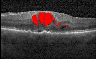
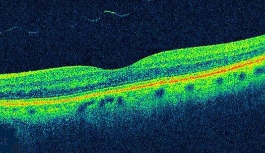
What are OCT Scans? Optical coherence tomography (OCT) is a non-invasive imaging method, which produces high-resolution volumetric histological images of tissue. To penetrate deep into biological

Read More

Finding Cysts Part Four: Seed Detection

A series of five articles on our Cysts Detection project using deep learning and Convolutional Neural Networks: 1) our cyst detection method; 2) the cyst denoising process; 3) the retinal layer segmentation; 4) the automatical seed-detection; 5) the final detection of the cysts. Our method is exceptionally successful at finding the cysts themselves and most of their area. Remarkable results are achieved even when using relatively small datasets in the training process.

Read More
Layer segmentation of the retina

Finding Cysts Part Three: Layer Segmentation

A series of five articles on our Cysts Detection project using deep learning and Convolutional Neural Networks: 1) our cyst detection method; 2) the cyst denoising process; 3) the retinal layer segmentation; 4) the automatical seed-detection; 5) the final detection of the cysts. Our method is exceptionally successful at finding the cysts themselves and most of their area. Remarkable results are achieved even when using relatively small datasets in the training process.

Read More
Denoising macular layers

Finding Cysts, Part Two: The Denoising Process

A series of five articles on our Cysts Detection project using deep learning and Convolutional Neural Networks: 1) our cyst detection method; 2) the cyst denoising process; 3) the retinal layer segmentation; 4) the automatical seed-detection; 5) the final detection of the cysts. Our method is exceptionally successful at finding the cysts themselves and most of their area. Remarkable results are achieved even when using relatively small datasets in the training process.

Read More

Automatic Detection of Macular Cysts

A series of five articles on our Cysts Detection project using deep learning and Convolutional Neural Networks: 1) our cyst detection method; 2) the cyst denoising process; 3) the retinal layer segmentation; 4) the automatical seed-detection; 5) the final detection of the cysts. Our method is exceptionally successful at finding the cysts themselves and most of their area. Remarkable results are achieved even when using relatively small datasets in the training process.

Read More

Get in touch

Please fill the following form and our experts will be happy to reply to you soon

Recent News

IBD Scoring – Clario, GI Reviewers and RSIP Vision Team Up

RSIP Neph Announces a Revolutionary Intra-op Solution for Partial Nephrectomy Surgeries

Announcement – RSIP Vision Presents Successful Preliminary Results from Clinical Study of 2D-to-3D Knee Bones Reconstruction

Announcement – New Urological AI Tool for 3D Reconstruction of the Ureter

All news
Upcoming Events
Stay informed for our next events
Subscribe to Our Magazines

Subscribe now and receive the Computer Vision News Magazine every month to your mailbox

 
Subscribe for free
Follow us
Linkedin Twitter Facebook Youtube

contact@rsipvision.com

Terms of Use

Privacy Policy

© All rights reserved to RSIP Vision 2023

Created by Shmulik

  • Our Work
    • title-1
      • Ophthalmology
      • Uncategorized
      • Ophthalmology
      • Pulmonology
      • Cardiology
      • Orthopedics
    • Title-2
      • Orthopedics
  • Success Stories
  • Insights
  • The company