Skip to content
  • Our Work
    • Fields
      • Cardiology
      • ENT
      • Gastro
      • Orthopedics
      • Ophthalmology
      • Pulmonology
      • Surgical
      • Urology
      • Other
    • Modalities
      • Endoscopy
      • Medical Segmentation
      • Microscopy
      • Ultrasound
  • Success Stories
  • Insights
    • Magazine
    • Upcoming Events
    • Webinars
    • Meetups
    • News
    • Blog
  • The company
    • About us
    • Careers
Menu
  • Our Work
    • Fields
      • Cardiology
      • ENT
      • Gastro
      • Orthopedics
      • Ophthalmology
      • Pulmonology
      • Surgical
      • Urology
      • Other
    • Modalities
      • Endoscopy
      • Medical Segmentation
      • Microscopy
      • Ultrasound
  • Success Stories
  • Insights
    • Magazine
    • Upcoming Events
    • Webinars
    • Meetups
    • News
    • Blog
  • The company
    • About us
    • Careers
Contact

Finding Cysts Part Four: Seed Detection

Seed Detection

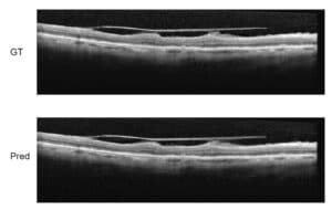
We’ve been working on automatically detecting Cystoid Macular Edema (CME) shown in Optical Coherence Tomography (OCT) images. CME  is a buildup of fluids in the eye near the macula of the eye.  OCT is one of the main modalities used to diagnose CME.
Our goal is to develop a system that can process large amounts of OCT images, automatically singling out images that show CME and quantify the cysts.
We have developed a multi-process method for more efficient automatic detection. The previous articles described the overall detection method, the denoising process, and layer segmentation. The next step is seed detection.
The goal of seed detection is to identify super-pixels in the scans that belong to a cyst. To do this, we will use machine learning methods with robust image processing features and build a classifier that can automatically detect cyst super-pixels in new images.

Super Pixels

Super-pixels are an increasingly popular computer vision concept where neighboring pixels are grouped into coherent units with perceptually shared meanings. Using this concept, a super-pixel carries much more meaning both geometrically and texturally than a single pixel. This allows us to create a meaningful classifier for super-pixels.
Our classifier will take into consideration the textural features of the super-pixel as well as its spatial location (which correlates to the distance of the area from the fovea) to determine whether the super-pixel belongs to a cyst or not.
As our features we use both statistics (skewness, kurtosis and more), spatial features (x,y coordinates) and various textural features (most prominent are the gabor filters, local binary patterns (LBP) and acutance).

Our Process

First we pre-process the image by cropping out the retina using the previously segmented RPE and ILM (see Layer Segmentation section for more detail). Next we segment the retina into super-pixels using the SLIC algorithm, which was introduced in the Denoising process.
From each super-pixel we extract the features mentioned before and train an ensemble of random forests combined with the adaBoost method to select the best features and cross-validate the results using the leave-one-out approach.

Estimating our Results

We had two manual annotations of the scans and due to the difficulty and subjectivity of defining a cyst have chosen to take a conservative approach and consider a proper “cyst” only cysts that were detected in both annotations.
In general we have found that using an ensemble of 32 forests is optimal with an average of 0.9243 accuracy, 0.92 sensitivity and 0.9253 specificity.
We  expect that by adding data from neighboring super-pixels to our classifier we will be able to get much better results.

Share

Share on linkedin
Share on twitter
Share on facebook

Main Field

Ophthalmology

RSIP Vision has developed countless projects in the field of ophthalmology for its clients. Computer vision with sophisticated ophthalmic imaging, measurement techniques and AI can yield better results: great precision, accurate diagnosis and best interventional treatment for many pathologies.

View Ophthalmology

Categories

  • Ophthalmology, RSIP Vision Learns

Related Content

Zoom-in-Net

Deep Learning in Ophthalmology

Classification and Segmentation of Dendritic cells

Classification and Segmentation of Dendritic Cells

Alzheimer's Disease - AD

Degenerative Diseases Detection in the Eye

Temporary pediatric strabismus in newborn baby

Image processing for pediatric strabismus

ROP - Vessel tortuosity in Retinopathy of Prematurity

ROP: Retinopathy of Prematurity

Eyelid Drooping - MRD1 and MRD2

Eyelid Drooping – Blepharoptosis

Show all

RSIP Vision

Field-tested software solutions and custom R&D, to power your next medical products with innovative AI and image analysis capabilities.

Read more about us

Get in touch

Please fill the following form and our experts will be happy to reply to you soon

Recent News

IBD Scoring – Clario, GI Reviewers and RSIP Vision Team Up

RSIP Neph Announces a Revolutionary Intra-op Solution for Partial Nephrectomy Surgeries

Announcement – RSIP Vision Presents Successful Preliminary Results from Clinical Study of 2D-to-3D Knee Bones Reconstruction

Announcement – New Urological AI Tool for 3D Reconstruction of the Ureter

All news
Upcoming Events
Stay informed for our next events
Subscribe to Our Magazines

Subscribe now and receive the Computer Vision News Magazine every month to your mailbox

 
Subscribe for free
Follow us
Linkedin Twitter Facebook Youtube

contact@rsipvision.com

Terms of Use

Privacy Policy

© All rights reserved to RSIP Vision 2023

Created by Shmulik

  • Our Work
    • title-1
      • Ophthalmology
      • Uncategorized
      • Ophthalmology
      • Pulmonology
      • Cardiology
      • Orthopedics
    • Title-2
      • Orthopedics
  • Success Stories
  • Insights
  • The company