Skip to content
  • Our Work
    • Fields
      • Cardiology
      • ENT
      • Gastro
      • Orthopedics
      • Ophthalmology
      • Pulmonology
      • Surgical
      • Urology
      • Other
    • Modalities
      • Endoscopy
      • Medical Segmentation
      • Microscopy
      • Ultrasound
  • Success Stories
  • Insights
    • Magazine
    • Upcoming Events
    • Webinars
    • Meetups
    • News
    • Blog
  • The company
    • About us
    • Careers
Menu
  • Our Work
    • Fields
      • Cardiology
      • ENT
      • Gastro
      • Orthopedics
      • Ophthalmology
      • Pulmonology
      • Surgical
      • Urology
      • Other
    • Modalities
      • Endoscopy
      • Medical Segmentation
      • Microscopy
      • Ultrasound
  • Success Stories
  • Insights
    • Magazine
    • Upcoming Events
    • Webinars
    • Meetups
    • News
    • Blog
  • The company
    • About us
    • Careers
Contact

Retinal Detachment Detection

Retinal detachment occurs when part of the retina detaches itself from the pigmented cell layer of the RPE, depriving itself of blood and nutrition: this condition causes blur and loss of vision.
Giant Retinal Tear
Giant Retinal Tear

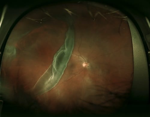
There are 2 types of detachment: either it is caused by fluids, which might be of lacrimal origins, gathering underneath the retina and lifting it; or it happens when the retina is pulled away together with the vitreous gel of the eye itself. The main reasons for the first might be retinal thinning, AMD or other pathologies, while the main reasons for the second might be excessive adhesion of retinal layers to contracting gel inside the aging eye, leads to a retinal tear from the RPE layer.

Retinal detachment is a medical emergency situation and needs to be treated immediately, generally by surgery, which fixes the retinal tears using photocoagulation (a laser treatment) or other kinds of procedures.

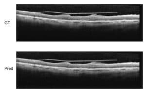
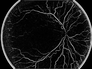
Detection of retinal detachment

Retinal detachment caused by fluids concentrating between the RPE and the inner layers of the retina. In some cases, fluids do not appear so clear in the OCT images and actually look grey, but still darker than the retinal layers. During the layers segmentation, the algorithm tracking the layers from left to right uses some optimization method.

Whenever we have a high curvature in the layer detection or a dark area above the lower layer, we must try to segment the cyst and by that we can give an estimation of the volume of the liquid found under the tear. In many cases where a cyst is found, the regular RPE to ILM measurement might give questionable results. But since we concentrate here in finding this edema, we are equipped with much better models for the pathology and can do a much better segmentation. The techniques that we will choose are a combination of various graph algorithms: either Bellman-Ford or Dijkstra will enable us to isolate the layers. Another segmentation algorithm, based on basic analysis, will tell us whether we have found a cyst or not. After that, we will be at ease to make the final retinal detachment segmentation using graph cuts, region growing or Otsu, an image processing binarization algorithm named after Nobuyuki Otsu, a Japanese mathematician.

Share

Share on linkedin
Share on twitter
Share on facebook

Main Field

Ophthalmology

RSIP Vision has developed countless projects in the field of ophthalmology for its clients. Computer vision with sophisticated ophthalmic imaging, measurement techniques and AI can yield better results: great precision, accurate diagnosis and best interventional treatment for many pathologies.

View Ophthalmology

Categories

  • Ophthalmology, RSIP Vision Learns

Related Content

Zoom-in-Net

Deep Learning in Ophthalmology

Classification and Segmentation of Dendritic cells

Classification and Segmentation of Dendritic Cells

Alzheimer's Disease - AD

Degenerative Diseases Detection in the Eye

Temporary pediatric strabismus in newborn baby

Image processing for pediatric strabismus

ROP - Vessel tortuosity in Retinopathy of Prematurity

ROP: Retinopathy of Prematurity

Eyelid Drooping - MRD1 and MRD2

Eyelid Drooping – Blepharoptosis

Show all

RSIP Vision

Field-tested software solutions and custom R&D, to power your next medical products with innovative AI and image analysis capabilities.

Read more about us

Get in touch

Please fill the following form and our experts will be happy to reply to you soon

Recent News

IBD Scoring – Clario, GI Reviewers and RSIP Vision Team Up

RSIP Neph Announces a Revolutionary Intra-op Solution for Partial Nephrectomy Surgeries

Announcement – RSIP Vision Presents Successful Preliminary Results from Clinical Study of 2D-to-3D Knee Bones Reconstruction

Announcement – New Urological AI Tool for 3D Reconstruction of the Ureter

All news
Upcoming Events
Stay informed for our next events
Subscribe to Our Magazines

Subscribe now and receive the Computer Vision News Magazine every month to your mailbox

 
Subscribe for free
Follow us
Linkedin Twitter Facebook Youtube

contact@rsipvision.com

Terms of Use

Privacy Policy

© All rights reserved to RSIP Vision 2023

Created by Shmulik

  • Our Work
    • title-1
      • Ophthalmology
      • Uncategorized
      • Ophthalmology
      • Pulmonology
      • Cardiology
      • Orthopedics
    • Title-2
      • Orthopedics
  • Success Stories
  • Insights
  • The company