Skip to content
  • Our Work
    • Fields
      • Cardiology
      • ENT
      • Gastro
      • Orthopedics
      • Ophthalmology
      • Pulmonology
      • Surgical
      • Urology
      • Other
    • Modalities
      • Endoscopy
      • Medical Segmentation
      • Microscopy
      • Ultrasound
  • Success Stories
  • Insights
    • Magazine
    • Upcoming Events
    • Webinars
    • Meetups
    • News
    • Blog
  • The company
    • About us
    • Careers
Menu
  • Our Work
    • Fields
      • Cardiology
      • ENT
      • Gastro
      • Orthopedics
      • Ophthalmology
      • Pulmonology
      • Surgical
      • Urology
      • Other
    • Modalities
      • Endoscopy
      • Medical Segmentation
      • Microscopy
      • Ultrasound
  • Success Stories
  • Insights
    • Magazine
    • Upcoming Events
    • Webinars
    • Meetups
    • News
    • Blog
  • The company
    • About us
    • Careers
Contact

Retinal images taken with mobile cameras

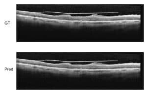
Portable cameras able to help ophthalmologists have been a desired solution for a long time. Among the reasons for the need of an additional device to support the eye physician, on top of available fixed devices, is the fact that sometimes patients who need the visit of  a doctor live in remote areas (or are housebound). In other hard to reach areas, there may be a total absence of any ophthalmologist able to collect medical information from patients. Initial retinal images taken with mobile cameras allow a first screening and first emergency decisions about the patient.

Retinal image with smartphone

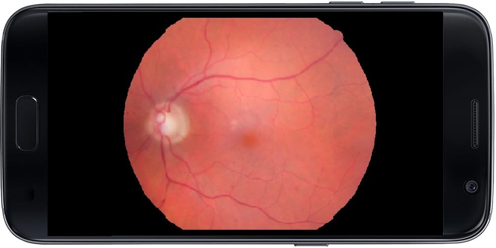
Together with telemedicine infrastructure, such retinal images taken with a mobile camera in remote places are transmitted to the ophthalmologist, who will thus be enabled to declare any pathology which can be assessed from the pictures. This is particularly useful, for instance, when screening cases of diabetic retinopathy: people with diabetes are at risk of developing diabetic retinopathy and therefore, need a regular screening with correct and timely diagnosis without the constraint of a long travel or needless waste of time, either for the eye care professional or for the patient: retinal images taken with mobile cameras are transmitted and analyzed remotely, regardless of distance.

Nowadays, when we have very good smartphone cameras, it is quite simple to add optics to the mobile device and look through the pupils to capture the retina. However, these settings present also several challenges: first and foremost, hand motion, eye motion and the system itself which is not stabilized like in the clinic. Next, the optics are not ideal and include lots of artifacts, like shadows and reflections, together with some mist or foggy effect that will reduce contrast and make everything look blurred. Light conditions might also be far from ideal.

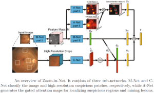
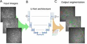
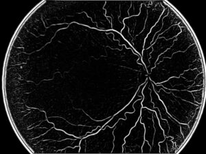
The solution to these challenges comes from image processing techniques. It is possible to identify and use automatic focus detection, to find the ideal focus for the image of the eye. The following step is to try and improve the image, finding the appropriate color transformation which offers the best result. Stitching of the images is also an essential process, since the effective field of view is very narrow, much narrower than regular fundus images. This is mainly due to shadows and reflections reducing sometimes more than 50% of the interested area. RSIP Vision has a very large experience in this stitching process: this enables us to take the best part of each image and stitch it in a montage of all the best parts, which will give a good panoramic view of the retina. This process can be done in the clinic, in better conditions than in the rural place where the original image was first taken. Also, processing power is probably much stronger on this side of the telemedicine channel than it can be on the smartphone or any other mobile device.

Share

Share on linkedin
Share on twitter
Share on facebook

Main Field

Ophthalmology

RSIP Vision has developed countless projects in the field of ophthalmology for its clients. Computer vision with sophisticated ophthalmic imaging, measurement techniques and AI can yield better results: great precision, accurate diagnosis and best interventional treatment for many pathologies.

View Ophthalmology

Categories

  • Ophthalmology, RSIP Vision Learns

Related Content

Zoom-in-Net

Deep Learning in Ophthalmology

Classification and Segmentation of Dendritic cells

Classification and Segmentation of Dendritic Cells

Alzheimer's Disease - AD

Degenerative Diseases Detection in the Eye

Temporary pediatric strabismus in newborn baby

Image processing for pediatric strabismus

ROP - Vessel tortuosity in Retinopathy of Prematurity

ROP: Retinopathy of Prematurity

Eyelid Drooping - MRD1 and MRD2

Eyelid Drooping – Blepharoptosis

Show all

RSIP Vision

Field-tested software solutions and custom R&D, to power your next medical products with innovative AI and image analysis capabilities.

Read more about us

Get in touch

Please fill the following form and our experts will be happy to reply to you soon

Recent News

IBD Scoring – Clario, GI Reviewers and RSIP Vision Team Up

RSIP Neph Announces a Revolutionary Intra-op Solution for Partial Nephrectomy Surgeries

Announcement – RSIP Vision Presents Successful Preliminary Results from Clinical Study of 2D-to-3D Knee Bones Reconstruction

Announcement – New Urological AI Tool for 3D Reconstruction of the Ureter

All news
Upcoming Events
Stay informed for our next events
Subscribe to Our Magazines

Subscribe now and receive the Computer Vision News Magazine every month to your mailbox

 
Subscribe for free
Follow us
Linkedin Twitter Facebook Youtube

contact@rsipvision.com

Terms of Use

Privacy Policy

© All rights reserved to RSIP Vision 2023

Created by Shmulik

  • Our Work
    • title-1
      • Ophthalmology
      • Uncategorized
      • Ophthalmology
      • Pulmonology
      • Cardiology
      • Orthopedics
    • Title-2
      • Orthopedics
  • Success Stories
  • Insights
  • The company