Skip to content
  • Our Work
    • Fields
      • Cardiology
      • ENT
      • Gastro
      • Orthopedics
      • Ophthalmology
      • Pulmonology
      • Surgical
      • Urology
      • Other
    • Modalities
      • Endoscopy
      • Medical Segmentation
      • Microscopy
      • Ultrasound
  • Success Stories
  • Insights
    • Magazine
    • Upcoming Events
    • Webinars
    • Meetups
    • News
    • Blog
  • The company
    • About us
    • Careers
Menu
  • Our Work
    • Fields
      • Cardiology
      • ENT
      • Gastro
      • Orthopedics
      • Ophthalmology
      • Pulmonology
      • Surgical
      • Urology
      • Other
    • Modalities
      • Endoscopy
      • Medical Segmentation
      • Microscopy
      • Ultrasound
  • Success Stories
  • Insights
    • Magazine
    • Upcoming Events
    • Webinars
    • Meetups
    • News
    • Blog
  • The company
    • About us
    • Careers
Contact

Diabetic retinopathy screening and microaneurysm detection

Diabetic Retinopathy (DR) is a leading cause of blindness, especially among adults and even more among the elderly segments of the population. It is associated with type 1 and type 2 diabetes. It occurs when high glucose levels in the blood damage blood vessels in the retinal area, leading to small leakages of blood, blood proteins, serum and blood fats.


Diabetic Retinal Screening

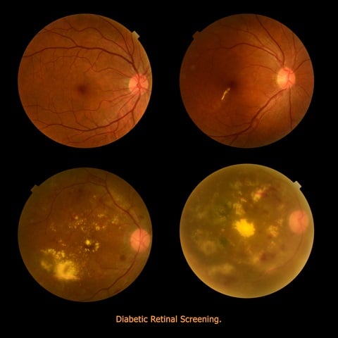
DR could be better treated if detected earlier. However, symptoms are limited and this often prevents a timely and appropriate treatment. Non-proliferative Diabetic Retinopathy (NPDR) is the earlier of the two stages of this pathology. At this stage, fats and other deposits cause micro aneurysms: spots on the walls of blood vessels in the retina. When these outpouch and leak, they cause swelling in the macula and in the nerve fibers. Capillaries may be obtruded and macula stops to work properly due to lack of oxygen, blurring the vision of the patient in the process.

Proliferative Diabetic Retinopathy (PDR) is a later stage of the disease. It occurs when many blood vessels close and then the retina tries to compensate by generating new blood vessels (neovascularization). However, these abnormal vessels do not supply the appropriate amount of blood to the retina. Scar tissues are stimulated by neovascularization, possibly provoking a retinal detachment. Another possible consequence is a damage to the optic nerve resulting in glaucoma.

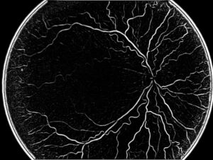
In terms of imaging, there are several ways to diagnose Diabetic Retinopathy: in the past, the most common method was a fluorescein test during angiography. This modality gives a video clip including the whole process, with fluorescein being visible in the arteries, getting absorbed in tissue and finally going back through the veins. Diabetic Retinopathy screening detects leaks of blood and other elements, giving evidence to the advancement of the disease.

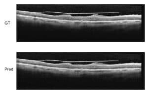
A more modern way to perform Diabetic Retinopathy screening is OCT, which offers the ophthalmologist a very precise angiography image without being invasive for the patient (no injection of substance), which eliminates many side-effects. A third method uses ultrasound to detect Diabetic Retinopathy symptoms: also B-scan ultrasonography enables the screening of blood vessels in the retinal area.

An automated algorithmic approach to detect and quantify Diabetic Retinopathy needs to fulfill several tasks: finding the blood leaks, detecting the growth of new capillaries, which indicate the presence of micro aneurysm. This is done by scanning carefully each pixel in the image, which is a time-consuming process when performed by a human, which justifies the quest for an automated procedure for microaneurysm detection.

This is a tricky problem to solve, since micro aneurysms look like small red dots, very similar to sections of blood vessels typically present in angiography images. Many steps are needed to clear up noise in these images. The first step is preprocessing, including boundary enhancement and more. Following that, we detect those suspected points using other filters like Hough Transform for circles together with other detectors.

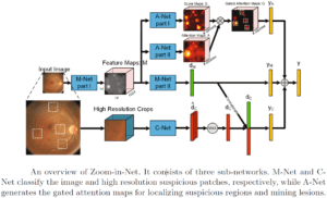
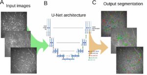
This methodology generates many false candidates (false positives) and the following step needs to use machine learning techniques to train a database of micro aneurysms. When enough data is collected, it is possible to use one of the known algorithms for the detection part. In this area, the use of deep learning is very efficient since we can limit the area and give the Convolutional Neural Networks (CNN) enough information to take the decision whether a suspected dot is a micro aneurysm or just a false alarm.

Share

Share on linkedin
Share on twitter
Share on facebook

Main Field

Ophthalmology

RSIP Vision has developed countless projects in the field of ophthalmology for its clients. Computer vision with sophisticated ophthalmic imaging, measurement techniques and AI can yield better results: great precision, accurate diagnosis and best interventional treatment for many pathologies.

View Ophthalmology

Categories

  • Ophthalmology, RSIP Vision Learns

Related Content

Zoom-in-Net

Deep Learning in Ophthalmology

Classification and Segmentation of Dendritic cells

Classification and Segmentation of Dendritic Cells

Alzheimer's Disease - AD

Degenerative Diseases Detection in the Eye

Temporary pediatric strabismus in newborn baby

Image processing for pediatric strabismus

ROP - Vessel tortuosity in Retinopathy of Prematurity

ROP: Retinopathy of Prematurity

Eyelid Drooping - MRD1 and MRD2

Eyelid Drooping – Blepharoptosis

Show all

RSIP Vision

Field-tested software solutions and custom R&D, to power your next medical products with innovative AI and image analysis capabilities.

Read more about us

Get in touch

Please fill the following form and our experts will be happy to reply to you soon

Recent News

IBD Scoring – Clario, GI Reviewers and RSIP Vision Team Up

RSIP Neph Announces a Revolutionary Intra-op Solution for Partial Nephrectomy Surgeries

Announcement – RSIP Vision Presents Successful Preliminary Results from Clinical Study of 2D-to-3D Knee Bones Reconstruction

Announcement – New Urological AI Tool for 3D Reconstruction of the Ureter

All news
Upcoming Events
Stay informed for our next events
Subscribe to Our Magazines

Subscribe now and receive the Computer Vision News Magazine every month to your mailbox

 
Subscribe for free
Follow us
Linkedin Twitter Facebook Youtube

contact@rsipvision.com

Terms of Use

Privacy Policy

© All rights reserved to RSIP Vision 2023

Created by Shmulik

  • Our Work
    • title-1
      • Ophthalmology
      • Uncategorized
      • Ophthalmology
      • Pulmonology
      • Cardiology
      • Orthopedics
    • Title-2
      • Orthopedics
  • Success Stories
  • Insights
  • The company