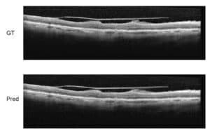
In a previous article, we talked about Geographic Atrophy segmentation in 2D images. This article focuses on how OCT images shed light on the development of Geographic Atrophy (GA) pathology. This spectral-domain optical coherence tomography (SD-OCT) modality with its 3D information enables a better volumetric and structural assessment of the GA, an advanced condition of Dry AMD (Age-related Macular Degeneration).
GA segmentation with SD-OCT images
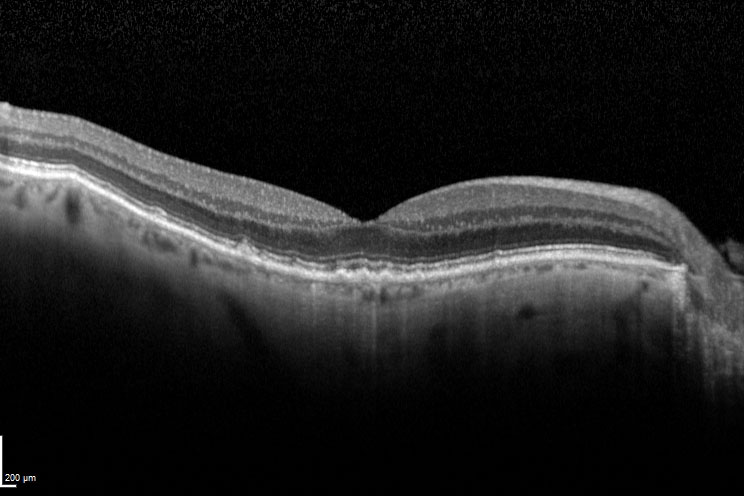
The SD-OCT shows several layers of the retina: the layer below the RPE (Retinal Pigment Epithelium) is the one where we can find evidence of Geographic Atrophy. Leading work in this area was done by researcher Luis de Sisternes, who published two subsequent articles on this subject. As it often happens, multi-modality imaging works very well on this problem: if you take FAF and IR fundus images, on top of OCT, you obtain a much better description of the GA, with much better analysis. As a matter of fact, RSIP Vision recommends to take images with several modalities: the collection of these images will be followed by a multi-modality registration task.
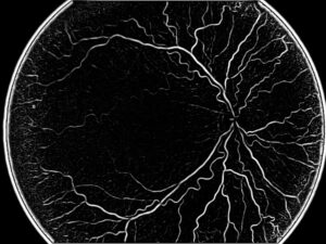
It is not a trivial matter to match these images, since they will be very different. One option is to take the OCT image and create a 2D angiography which will facilitate the registration of the other modalities. Registration needs to use feature point or mutual information techniques because of the different appearance of the images. In mutual information, we do not look at the specific information of the pixels in order to do the calculations. Rather, we consider the statistics of every image and this enables us to detect areas where greyscales are very different and even opposite. On the other hand, when we use key points, we can choose them to be robust enough for all different modalities. For example, a bifurcation will appear differently in FAF and in OCT, but the bifurcation is still the same: if this point is correctly detected, it becomes a very robust anchor point for the registration.
Did you read our other articles about image processing in ophthalmology?
 Ophthalmology