Skip to content
  • Our Work
    • Fields
      • Cardiology
      • ENT
      • Gastro
      • Orthopedics
      • Ophthalmology
      • Pulmonology
      • Surgical
      • Urology
      • Other
    • Modalities
      • Endoscopy
      • Medical Segmentation
      • Microscopy
      • Ultrasound
  • Success Stories
  • Insights
    • Magazine
    • Upcoming Events
    • Webinars
    • Meetups
    • News
    • Blog
  • The company
    • About us
    • Careers
Menu
  • Our Work
    • Fields
      • Cardiology
      • ENT
      • Gastro
      • Orthopedics
      • Ophthalmology
      • Pulmonology
      • Surgical
      • Urology
      • Other
    • Modalities
      • Endoscopy
      • Medical Segmentation
      • Microscopy
      • Ultrasound
  • Success Stories
  • Insights
    • Magazine
    • Upcoming Events
    • Webinars
    • Meetups
    • News
    • Blog
  • The company
    • About us
    • Careers
Contact

Measurement of Retinal Thickness

Retinal thickness is a key measurement used to assess the health of the retina and whether it needs any treatment. Thickness measure of a patient’s retina should ideally fall within a range, which varies according to the region of the eye.  For example, the retina is typically thinner in the center, in the foveal area.

Figures can be compared to optimal ranges or to data from the same patient over time: thinning retina can be the consequence of aging or of retinal disorders which might or might not need to be treated. In particular, the thickness, coherence and curvature of the retina layers have been shown to be related to a range of different retinopathies.

Retinal thickness
A case of curvature of the retina layer, which may suggest a retinopathy

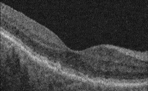
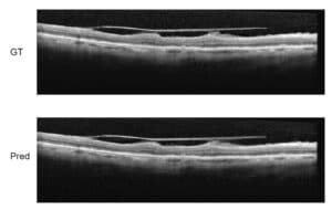
Across various definitions, retinal thickness is calculated as the distance between the edges of the retina: in this project we were asked by our client to measure the ILM to RPE total retinal thickness, where the ILM is the internal limiting membrane and the RPE is the retinal pigment epithelium.

We can measure the thickness of the retina from OCT (Optical Coherence Tomography) scans. These scans are 2D slices which need to be combined to create a 3D view which can be used by the medical professionals. Experienced ophthalmologists are able to manually identify ILM and RPE lines on OCT scans and their assessment of retinal thickness is reliable: however, doing that in a huge number of OCT slices is a very long and tedious activity, hence the need for an automatic measurement of retinal thickness in the areas of interest using computerized image processing algorithms calculating the distance of ILM to RPE.

The measurement of retinal thickness is generally performed on high-resolution OCT scans, but our client needed a more sophisticated solution able to give a correct measurement even when OCT is done with older, slower or otherwise less performing equipment, in order to allow ophthalmologists to still provide a correct diagnostics of the status of the retina. Of course, input of lower quality needs more sophisticated algorithms to compensate for missing or incomplete data in the OCT scans.

Advanced optimization methods, borrowed from graph theory – one of the active and fruitful fields in mathematics – helped us to solve the complex challenge by representing the problem as a graph and allowing to solve it over the complete slice (global optimization) through mathematical theorems, all within reasonable processing time.

The output provided by our software on lower quality devices was compared by our client with output given by better quality equipment and the results were overall equivalent, notwithstanding the challenging conditions. The software application specifically developed by RSIP Vision was found exceptionally reliable: it was therefore adopted by our client and is currently used on OCT equipment to perform automatic measurement of retinal thickness.

ETDRS Grid for OCT Retina Measurement
Our software determines the thickness of the retina in each of its 9 fields as defined by the ETDRS grid for measuring eye health (used in the Age Related Eye Disease Study – AREDS)

Share

Share on linkedin
Share on twitter
Share on facebook

Main Field

Ophthalmology

RSIP Vision has developed countless projects in the field of ophthalmology for its clients. Computer vision with sophisticated ophthalmic imaging, measurement techniques and AI can yield better results: great precision, accurate diagnosis and best interventional treatment for many pathologies.

View Ophthalmology

Categories

  • Ophthalmology, RSIP Vision Learns

Related Content

Zoom-in-Net

Deep Learning in Ophthalmology

Classification and Segmentation of Dendritic cells

Classification and Segmentation of Dendritic Cells

Alzheimer's Disease - AD

Degenerative Diseases Detection in the Eye

Temporary pediatric strabismus in newborn baby

Image processing for pediatric strabismus

ROP - Vessel tortuosity in Retinopathy of Prematurity

ROP: Retinopathy of Prematurity

Eyelid Drooping - MRD1 and MRD2

Eyelid Drooping – Blepharoptosis

Show all

RSIP Vision

Field-tested software solutions and custom R&D, to power your next medical products with innovative AI and image analysis capabilities.

Read more about us

Get in touch

Please fill the following form and our experts will be happy to reply to you soon

Recent News

IBD Scoring – Clario, GI Reviewers and RSIP Vision Team Up

RSIP Neph Announces a Revolutionary Intra-op Solution for Partial Nephrectomy Surgeries

Announcement – RSIP Vision Presents Successful Preliminary Results from Clinical Study of 2D-to-3D Knee Bones Reconstruction

Announcement – New Urological AI Tool for 3D Reconstruction of the Ureter

All news
Upcoming Events
Stay informed for our next events
Subscribe to Our Magazines

Subscribe now and receive the Computer Vision News Magazine every month to your mailbox

 
Subscribe for free
Follow us
Linkedin Twitter Facebook Youtube

contact@rsipvision.com

Terms of Use

Privacy Policy

© All rights reserved to RSIP Vision 2023

Created by Shmulik

  • Our Work
    • title-1
      • Ophthalmology
      • Uncategorized
      • Ophthalmology
      • Pulmonology
      • Cardiology
      • Orthopedics
    • Title-2
      • Orthopedics
  • Success Stories
  • Insights
  • The company