Skip to content
  • Our Work
    • Fields
      • Cardiology
      • ENT
      • Gastro
      • Orthopedics
      • Ophthalmology
      • Pulmonology
      • Surgical
      • Urology
      • Other
    • Modalities
      • Endoscopy
      • Medical Segmentation
      • Microscopy
      • Ultrasound
  • Success Stories
  • Insights
    • Magazine
    • Upcoming Events
    • Webinars
    • Meetups
    • News
    • Blog
  • The company
    • About us
    • Careers
Menu
  • Our Work
    • Fields
      • Cardiology
      • ENT
      • Gastro
      • Orthopedics
      • Ophthalmology
      • Pulmonology
      • Surgical
      • Urology
      • Other
    • Modalities
      • Endoscopy
      • Medical Segmentation
      • Microscopy
      • Ultrasound
  • Success Stories
  • Insights
    • Magazine
    • Upcoming Events
    • Webinars
    • Meetups
    • News
    • Blog
  • The company
    • About us
    • Careers
Contact

Retina Montage Software

The Need for Retina Montage:

Retinal imaging is one of the oldest and most reliable techniques to examine the retina to analyse its status and possibly detect existing disorders. The principle is very simple: a regular camera with proper optics and enough light can look through the pupil and see the fundus of the eye.

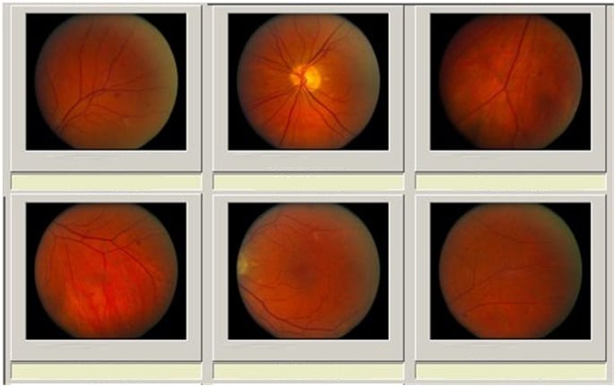
Unfortunately, the field of each single view is limited: the solution used by ophthalmologists is to ask patients to look up, down and on the sides in order to collect images covering all the area of the retina. After doing that, images need to be combined together into a single mosaic view, offering a general panorama of the retina. This way of providing the eye care professional a larger field of view is generally called retinal image montage or panorama.

Retina Panorama
RSIP Vision’s stitching technology enables the ophthalmologist to analyze the patient’s retinal vascular tree through an efficient and noninvasive process

Stitch Together Multiple Fundus Images:

The first step of mosaicing eye fundus images can be done in at least two ways: one way scans the eye following a very strict scheme, but this is not very comfortable for the ophthalmologist and the patient. The other way is less strict about the scheme while taking enough images of the different areas of the retina. The angles can be chosen arbitrarily: provided all the area of interest is covered by the images, we are able to adapt images taken from different angles and merge them within a single registration.
Retina Panorama
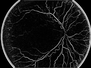
The following step to achieve retina montage requires to combine those images together into a single montage of the retina, solving a sort of puzzle. Overlapping areas are generally not large, hence giving little information on the rear parts of the retina, where registered images might provide only one significant vessel. An additional challenge is given by the fact that the margins of the images, where the overlapping needs to be found, have different lighting conditions and are often corrupted: this results in blurry and grey margins, calling for color corrections in this areas before stitching the image into a single retinal montage. This enables to gradually fuse multiple retinal images, when moving from one image to the other in the finally aligned mosaic.

Image Stitching Software:

It cannot be stressed enough how accurate the combination of these multiple retinal images needs to be, including obtaining confirmation that the stitching solution found is valid. Any solution which includes errors will deceive the ophthalmologist by giving him a wrong panoramic view. Being the eye in spherical form, this appears as a three-dimensional problem: only if you solve the puzzle on a 3D sphere, images will match accurately. Images combined on a 2D plane will not match: the incorrect alignments will result in duplication of arteries and similar incongruities.

Our Retina Montage Software Solution:

RSIP Vision uses very advanced technologies to perform this ophthalmology software task, including graph theory techniques to obtain the best optimization of retina montage. Our in-house developed montaging method uses very sophisticated algorithms, enabling us to emphasize information which is sometimes very dull and noisy. The final result is the generation of a retinal montage displaying all the information needed to enable the ophthalmologist to give optimal diagnostic accuracy of the fundus of the eyes.

Share

Share on linkedin
Share on twitter
Share on facebook

Main Field

Ophthalmology

RSIP Vision has developed countless projects in the field of ophthalmology for its clients. Computer vision with sophisticated ophthalmic imaging, measurement techniques and AI can yield better results: great precision, accurate diagnosis and best interventional treatment for many pathologies.

View Ophthalmology

Categories

  • Ophthalmology, RSIP Vision Learns

Related Content

Zoom-in-Net

Deep Learning in Ophthalmology

Classification and Segmentation of Dendritic cells

Classification and Segmentation of Dendritic Cells

Alzheimer's Disease - AD

Degenerative Diseases Detection in the Eye

Temporary pediatric strabismus in newborn baby

Image processing for pediatric strabismus

ROP - Vessel tortuosity in Retinopathy of Prematurity

ROP: Retinopathy of Prematurity

Eyelid Drooping - MRD1 and MRD2

Eyelid Drooping – Blepharoptosis

Show all

RSIP Vision

Field-tested software solutions and custom R&D, to power your next medical products with innovative AI and image analysis capabilities.

Read more about us

Get in touch

Please fill the following form and our experts will be happy to reply to you soon

Recent News

IBD Scoring – Clario, GI Reviewers and RSIP Vision Team Up

RSIP Neph Announces a Revolutionary Intra-op Solution for Partial Nephrectomy Surgeries

Announcement – RSIP Vision Presents Successful Preliminary Results from Clinical Study of 2D-to-3D Knee Bones Reconstruction

Announcement – New Urological AI Tool for 3D Reconstruction of the Ureter

All news
Upcoming Events
Stay informed for our next events
Subscribe to Our Magazines

Subscribe now and receive the Computer Vision News Magazine every month to your mailbox

 
Subscribe for free
Follow us
Linkedin Twitter Facebook Youtube

contact@rsipvision.com

Terms of Use

Privacy Policy

© All rights reserved to RSIP Vision 2023

Created by Shmulik

  • Our Work
    • title-1
      • Ophthalmology
      • Uncategorized
      • Ophthalmology
      • Pulmonology
      • Cardiology
      • Orthopedics
    • Title-2
      • Orthopedics
  • Success Stories
  • Insights
  • The company