Skip to content
  • Our Work
    • Fields
      • Cardiology
      • ENT
      • Gastro
      • Orthopedics
      • Ophthalmology
      • Pulmonology
      • Surgical
      • Urology
      • Other
    • Modalities
      • Endoscopy
      • Medical Segmentation
      • Microscopy
      • Ultrasound
  • Success Stories
  • Insights
    • Magazine
    • Upcoming Events
    • Webinars
    • Meetups
    • News
    • Blog
  • The company
    • About us
    • Careers
Menu
  • Our Work
    • Fields
      • Cardiology
      • ENT
      • Gastro
      • Orthopedics
      • Ophthalmology
      • Pulmonology
      • Surgical
      • Urology
      • Other
    • Modalities
      • Endoscopy
      • Medical Segmentation
      • Microscopy
      • Ultrasound
  • Success Stories
  • Insights
    • Magazine
    • Upcoming Events
    • Webinars
    • Meetups
    • News
    • Blog
  • The company
    • About us
    • Careers
Contact

ROP: Retinopathy of Prematurity

Retinopathy of prematurity (ROP) is a leading cause of blindness in infants. ROP (or Terry syndrome) is a disease of the eye affecting prematurely-born, low birthweight infants having received intensive neonatal care which includes oxygen therapy. Oxygen toxicity causes abnormal growth of retinal blood vessels. These vascular changes are described as plus disease and are defined as abnormal dilation and tortuosity of the blood vessels, which are fragile and can leak and scar the retina, potentially leading to retinal detachment. The longer retinal detachment goes untreated, the greater the risk of permanent vision loss in the affected eye. Retinopathy of prematurity is an emergency condition.
ROP - Vessel tortuosity in Retinopathy of Prematurity
ROP – Vessel tortuosity in Retinopathy of Prematurity fundus image

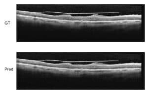
ROP shows no external signs or symptoms. The only way to detect it is through an eye examination by an ophthalmologist. Telemedicine and automated image analysis have the potential to improve access to eye care in less developed and accessible regions, boosting the quality of ROP care.

Computer-based Imaging for Retinopathy of Prematurity diagnosis

Computer-based image analysis has the potential to produce quantifiable, objective measurements.

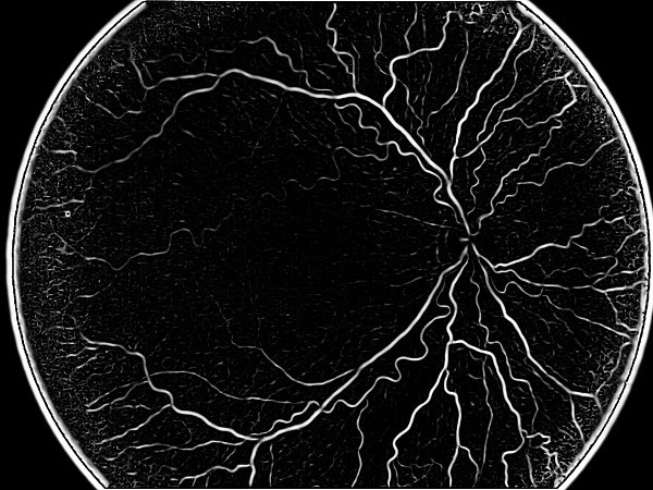
Most approaches for diagnosis include segmentation of retinal vessels and extraction from fundus images of features that account for tortuosity, dilation (diameter) and integrated curvature (sum of angles) for each vessel.

These approaches differ in the various methods used to process the images in order to receive quantifiable values. The first step is pre-processing the images: removing saturation noise by equalizing the histogram of the image and thresholding it to form a binary image. The global threshold value should be adjusted according to the efficiency of thresholding. It is necessary to omit the Optic Disc boundaries before proceeding toward feature extraction. A circular mask can do this job due to the circular morphology of the Optic Disc. Since most ROP features are found close to the optic disc, it is important to define the diagnostic regions at some radius around it to compute features that are relevant to the assessment of the pathology. Next, the vessel centerlines are extracted and a vasculature tree is constructed.

Vessel segmentation approaches are based on algorithmic techniques like matched filter response, multiscale image analysis, morphological segmentation and region growing. For instance, matched filter response measures responses for a binary image with a set of templates in different orientations, based on the assumption that vessels are aligned over the vertical axis and vessels gradients are symmetrical. The response is the outcome of convolution and only the maximum response for each pixel, which indicates of the best match, is retained. Finally, vessel post-processing takes place and measurements are taken for all desired features.

RSIP Vision has a very large portfolio of ophthalmology image analysis projects. This R&D and consulting experience can certainly contribute to the success of your project. Talk about it with our engineers!

Contact us now

Share

Share on linkedin
Share on twitter
Share on facebook

Main Field

Ophthalmology

RSIP Vision has developed countless projects in the field of ophthalmology for its clients. Computer vision with sophisticated ophthalmic imaging, measurement techniques and AI can yield better results: great precision, accurate diagnosis and best interventional treatment for many pathologies.

View Ophthalmology

Categories

  • Ophthalmology, RSIP Vision Learns

Related Content

Zoom-in-Net

Deep Learning in Ophthalmology

Classification and Segmentation of Dendritic cells

Classification and Segmentation of Dendritic Cells

Alzheimer's Disease - AD

Degenerative Diseases Detection in the Eye

Temporary pediatric strabismus in newborn baby

Image processing for pediatric strabismus

Eyelid Drooping - MRD1 and MRD2

Eyelid Drooping – Blepharoptosis

Fundus image

Retinal images enhancement

Show all

RSIP Vision

Field-tested software solutions and custom R&D, to power your next medical products with innovative AI and image analysis capabilities.

Read more about us

Get in touch

Please fill the following form and our experts will be happy to reply to you soon

Recent News

IBD Scoring – Clario, GI Reviewers and RSIP Vision Team Up

RSIP Neph Announces a Revolutionary Intra-op Solution for Partial Nephrectomy Surgeries

Announcement – RSIP Vision Presents Successful Preliminary Results from Clinical Study of 2D-to-3D Knee Bones Reconstruction

Announcement – New Urological AI Tool for 3D Reconstruction of the Ureter

All news
Upcoming Events
Stay informed for our next events
Subscribe to Our Magazines

Subscribe now and receive the Computer Vision News Magazine every month to your mailbox

 
Subscribe for free
Follow us
Linkedin Twitter Facebook Youtube

contact@rsipvision.com

Terms of Use

Privacy Policy

© All rights reserved to RSIP Vision 2023

Created by Shmulik

  • Our Work
    • title-1
      • Ophthalmology
      • Uncategorized
      • Ophthalmology
      • Pulmonology
      • Cardiology
      • Orthopedics
    • Title-2
      • Orthopedics
  • Success Stories
  • Insights
  • The company