Skip to content
  • Our Work
    • Fields
      • Cardiology
      • ENT
      • Gastro
      • Orthopedics
      • Ophthalmology
      • Pulmonology
      • Surgical
      • Urology
      • Other
    • Modalities
      • Endoscopy
      • Medical Segmentation
      • Microscopy
      • Ultrasound
  • Success Stories
  • Insights
    • Magazine
    • Upcoming Events
    • Webinars
    • Meetups
    • News
    • Blog
  • The company
    • About us
    • Careers
Menu
  • Our Work
    • Fields
      • Cardiology
      • ENT
      • Gastro
      • Orthopedics
      • Ophthalmology
      • Pulmonology
      • Surgical
      • Urology
      • Other
    • Modalities
      • Endoscopy
      • Medical Segmentation
      • Microscopy
      • Ultrasound
  • Success Stories
  • Insights
    • Magazine
    • Upcoming Events
    • Webinars
    • Meetups
    • News
    • Blog
  • The company
    • About us
    • Careers
Contact

Optical Coherence Tomography Angiography

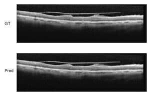
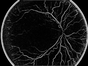
Optical coherence tomography angiography (OCTA) is a method which uses OCT to produce high-resolution volumetric images of the vascular tree in the eye. It has the advantage of being a non-invasive procedure. It is quick, and produces high resolution 3-dimensional histological images.

Optical Coherence Tomography Angiography

With OCTA a clear structure of the blood flow in the eye can be seen, which enables a better diagnosis of pathologies such as age-related macular degeneration, vein occlusion, and glaucoma. To create a map of the blood flow, OCTA compares sequential differences in the back-scattering of the light reflected from a specific cross-section of the eye. To obtain high resolution images, OCTA requires high-speed imaging. Due to the technical limitation of OCT machines, however, only a narrow field of view can be acquired as such.

An improvement in the amount of information obtained from OCTA is achieved over other methods such as Fluorescein angiography (FA). In the later, a fluorescein dye is injected to blood circulation to increase contrast during imaging when illuminated by blue light.

The procedure of FA is invasive, long (10-30 minutes) and prone to measurement error. Moreover, using dye in FA obscures the detection of lesions and diagnosis of their extent and depth, and does not allow easy segmentation of the different layers in the retina. In contrast, OCTA avoids this.

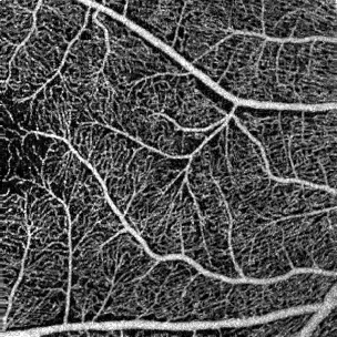
A further improvement in resolution of OCTA can be achieved by employing phase-contrast OCTA. Phase-contrast OCTA improves the resolution by at least an order of magnitude over FA and time-domain OCT which achieves sub-micrometer resolution. The way it works is by splitting a low coherence beam into two using a prism, the splat beams then pass through the tissue in two close paths and recombine upon exiting. All differences in phase between the two beams are translated into changes in the polarization of the signal which are later translated into phase-contrast OCT images.

Three-dimensional blood flow can then be visualized in OCTA by detecting regions of differential phase variance in multiple adjacent two-dimensional scans (b-scans) of individual slices of the retina. This allows physicians to quickly and accurately detect pathologies related to vascularization in a non-invasive manner which is practically free of side effects and clinical hazards.

RSIP Vision has vast experience in OCTA, formulating processes which stitch multiple images together, increase image information and clarity.

Share

Share on linkedin
Share on twitter
Share on facebook

Main Field

Ophthalmology

RSIP Vision has developed countless projects in the field of ophthalmology for its clients. Computer vision with sophisticated ophthalmic imaging, measurement techniques and AI can yield better results: great precision, accurate diagnosis and best interventional treatment for many pathologies.

View Ophthalmology

Categories

  • Ophthalmology, RSIP Vision Learns

Related Content

Zoom-in-Net

Deep Learning in Ophthalmology

Classification and Segmentation of Dendritic cells

Classification and Segmentation of Dendritic Cells

Alzheimer's Disease - AD

Degenerative Diseases Detection in the Eye

Temporary pediatric strabismus in newborn baby

Image processing for pediatric strabismus

ROP - Vessel tortuosity in Retinopathy of Prematurity

ROP: Retinopathy of Prematurity

Eyelid Drooping - MRD1 and MRD2

Eyelid Drooping – Blepharoptosis

Show all

RSIP Vision

Field-tested software solutions and custom R&D, to power your next medical products with innovative AI and image analysis capabilities.

Read more about us

Get in touch

Please fill the following form and our experts will be happy to reply to you soon

Recent News

IBD Scoring – Clario, GI Reviewers and RSIP Vision Team Up

RSIP Neph Announces a Revolutionary Intra-op Solution for Partial Nephrectomy Surgeries

Announcement – RSIP Vision Presents Successful Preliminary Results from Clinical Study of 2D-to-3D Knee Bones Reconstruction

Announcement – New Urological AI Tool for 3D Reconstruction of the Ureter

All news
Upcoming Events
Stay informed for our next events
Subscribe to Our Magazines

Subscribe now and receive the Computer Vision News Magazine every month to your mailbox

 
Subscribe for free
Follow us
Linkedin Twitter Facebook Youtube

contact@rsipvision.com

Terms of Use

Privacy Policy

© All rights reserved to RSIP Vision 2023

Created by Shmulik

  • Our Work
    • title-1
      • Ophthalmology
      • Uncategorized
      • Ophthalmology
      • Pulmonology
      • Cardiology
      • Orthopedics
    • Title-2
      • Orthopedics
  • Success Stories
  • Insights
  • The company