Skip to content
  • Our Work
    • Fields
      • Cardiology
      • ENT
      • Gastro
      • Orthopedics
      • Ophthalmology
      • Pulmonology
      • Surgical
      • Urology
      • Other
    • Modalities
      • Endoscopy
      • Medical Segmentation
      • Microscopy
      • Ultrasound
  • Success Stories
  • Insights
    • Magazine
    • Upcoming Events
    • Webinars
    • Meetups
    • News
    • Blog
  • The company
    • About us
    • Careers
Menu
  • Our Work
    • Fields
      • Cardiology
      • ENT
      • Gastro
      • Orthopedics
      • Ophthalmology
      • Pulmonology
      • Surgical
      • Urology
      • Other
    • Modalities
      • Endoscopy
      • Medical Segmentation
      • Microscopy
      • Ultrasound
  • Success Stories
  • Insights
    • Magazine
    • Upcoming Events
    • Webinars
    • Meetups
    • News
    • Blog
  • The company
    • About us
    • Careers
Contact

Retinal images enhancement

Image enhancement of retinal structures has the potential to facilitate diagnosis of several eye diseases. Retinal disease diagnosis and monitoring often requires very delicate analysis that can be only accomplished with appropriate resources, which include Fundus camera or OCT device with high resolution, as well as physician’s expertise and in many cases also image enhancements. These delicate analysis include segmentation, measurement and quantification, the accuracy of which relies on the quality of the input image. Image quality is affected by factors such as medial opacity, defocus or presence of artifact. Retinal image enhancement techniques are used to improve contrast or to brighten an image.

Fundus image


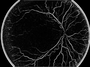
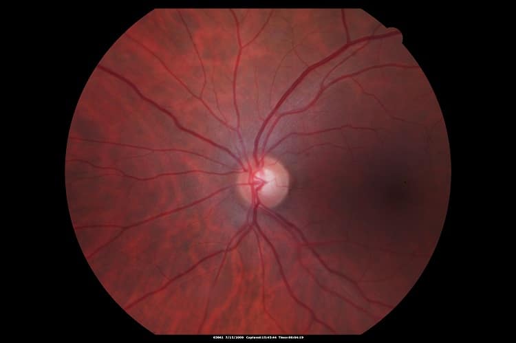
Fundus images enhancement

It is highly recommended to use image histogram stretching techniques in fundus images. Information about the distribution of grey levels in a specific image is provided by the image histogram. In an exceptionally dark or bright image, the grey level would be clustered to the extremes of the histogram, but in a well-contrasted image these levels would be well spread over a wide range. In low contrast image, the solution is given by histogram stretching algorithms: these algorithms are able to distribute grey levels more equally across the range, according to specific user-defined equations, and therefore produce an image with greater contrast than the original. Histogram equalization is a basic automated process, which aims at making the histogram uniformly distributed. When using an adaptive Histogram Equalization method, the image is divided to sub-images, each having a sub-histogram which is then stretched. The neighboring sub-images are stitched together using bilinear interpolation to eliminate the boundaries between them.

Other retinal images enhancement methods such as filtering make the image more suitable for use: one of these filtering methods is averaging, that is able to reduce noise; another filtering method involves boundaries enhancement.


OCT enhancement

OCT is extremely useful in assessing optic nerve damages in glaucoma, retinal detachment and macular edema. Evaluation of these pathologies requires assessment of changes over time, which might be very hard to tell in some images. OCT images are subject to various distortions that introduce artifacts in the OCT cross-sections such as floaters, saccades and speckle noise.

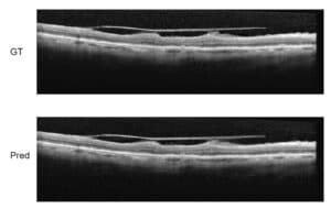
OCT retinal image enhancement methods are different from those which we have seen giving such good results on fundus images: signal averaging is one of the most important and effective techniques to improve OCT image quality. This technique has practical limitations due to the inability of patients to maintain fixation during examination and it requires hardware eye-tracking, which is not necessarily available in every clinic using OCT. Due to eye movement (eye saccades, blinks and patient’s movement) there is a limitation on the number of frames that can be recorded and averaged.

For that reason, we recommend using an enhancement method by virtually averaging OCT signal (no need for eye tracking). The technique uses Gaussian random distribution for each sampling voxel on a 3x3x3 cube of neighboring voxels; this is repeated an appropriate amount of times such as ensures the optimization of the averaging result.

Both signal to noise ratio (SNR) and contrast to noise ratio (CNR) are improved by this method: these parameters indicate that the non-averaged non-tracking OCT image quality is significantly better.

Another denoising technique consists in a locally adaptive denoising algorithm using a combination of the Double-Density Discrete Wavelet Transform (DDDWT) and the Dual-Tree Complex Wavelet Transform (DTCWT), useful to reduce speckle noise in OCT images of the retina. The algorithm overcomes the limitations of commonly used multiple frame averaging technique, improving SNR.

RSIP Vision has been at the forefront of these retinal images enhancement techniques for several decades. Coupled with an excellent knowledge of the eye’s physiology, our expertise will have an essential role in the success of your ophthalmology software. Contact our consultants to find out what we can do for you!

Share

Share on linkedin
Share on twitter
Share on facebook

Main Field

Ophthalmology

RSIP Vision has developed countless projects in the field of ophthalmology for its clients. Computer vision with sophisticated ophthalmic imaging, measurement techniques and AI can yield better results: great precision, accurate diagnosis and best interventional treatment for many pathologies.

View Ophthalmology

Categories

  • Ophthalmology, RSIP Vision Learns

Related Content

Zoom-in-Net

Deep Learning in Ophthalmology

Classification and Segmentation of Dendritic cells

Classification and Segmentation of Dendritic Cells

Alzheimer's Disease - AD

Degenerative Diseases Detection in the Eye

Temporary pediatric strabismus in newborn baby

Image processing for pediatric strabismus

ROP - Vessel tortuosity in Retinopathy of Prematurity

ROP: Retinopathy of Prematurity

Eyelid Drooping - MRD1 and MRD2

Eyelid Drooping – Blepharoptosis

Show all

RSIP Vision

Field-tested software solutions and custom R&D, to power your next medical products with innovative AI and image analysis capabilities.

Read more about us

Get in touch

Please fill the following form and our experts will be happy to reply to you soon

Recent News

IBD Scoring – Clario, GI Reviewers and RSIP Vision Team Up

RSIP Neph Announces a Revolutionary Intra-op Solution for Partial Nephrectomy Surgeries

Announcement – RSIP Vision Presents Successful Preliminary Results from Clinical Study of 2D-to-3D Knee Bones Reconstruction

Announcement – New Urological AI Tool for 3D Reconstruction of the Ureter

All news
Upcoming Events
Stay informed for our next events
Subscribe to Our Magazines

Subscribe now and receive the Computer Vision News Magazine every month to your mailbox

 
Subscribe for free
Follow us
Linkedin Twitter Facebook Youtube

contact@rsipvision.com

Terms of Use

Privacy Policy

© All rights reserved to RSIP Vision 2023

Created by Shmulik

  • Our Work
    • title-1
      • Ophthalmology
      • Uncategorized
      • Ophthalmology
      • Pulmonology
      • Cardiology
      • Orthopedics
    • Title-2
      • Orthopedics
  • Success Stories
  • Insights
  • The company