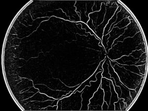
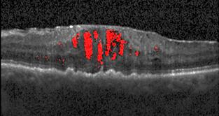
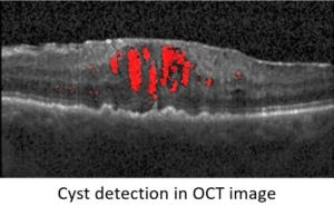
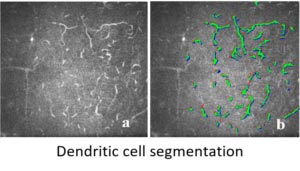
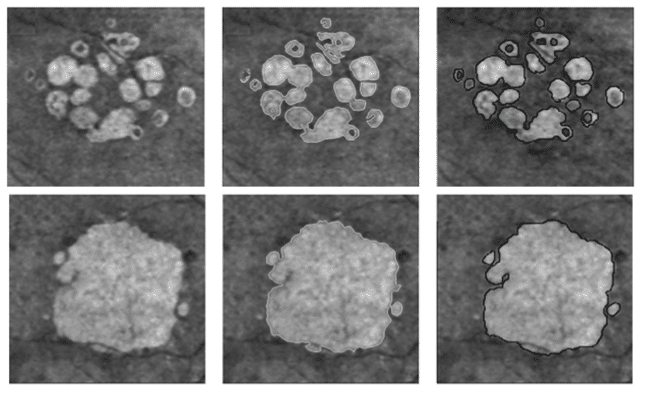
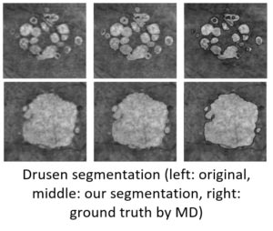
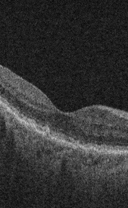
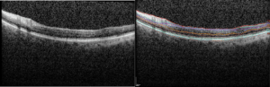
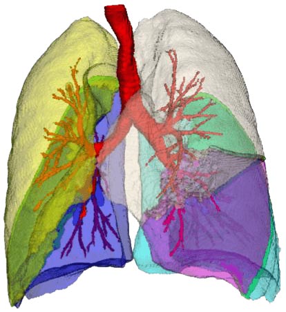
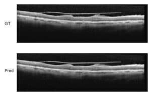
Super-Resolution in OCT images
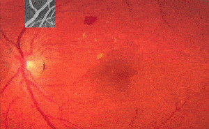
OCT images are frequently used to demonstrate the retina and understand the different pathologies and decide the diagnostics and help with procedure planning. In medical imaging, especially in radiology, deep learning has been used in recent years to gain super-resolution. On one hand, it is possible to scan faster and more efficiently at lower resolutions. On the other hand, quality scans provide added benefits. Super-resolution is a class of techniques in image processing that enhance the resolution of an imaging system, making images sharper and clearer. By improving the quality