Deep Learning in Ophthalmology
Recent works suggest novel deep learning tools for detection, segmentation and characterization of eye disorders. Accurate segmentation of retinal fundus lesions and anomalies in imaging
Recent works suggest novel deep learning tools for detection, segmentation and characterization of eye disorders. Accurate segmentation of retinal fundus lesions and anomalies in imaging
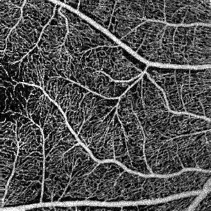
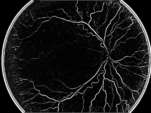
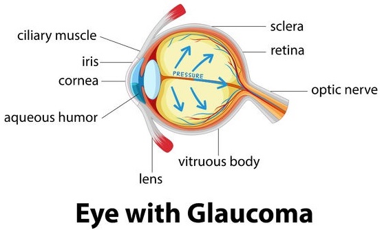
Modern imaging knows how to capture an image to see through the eye tissue transparency and inspect retina, vasculature and neural tissue: this phenomenon is
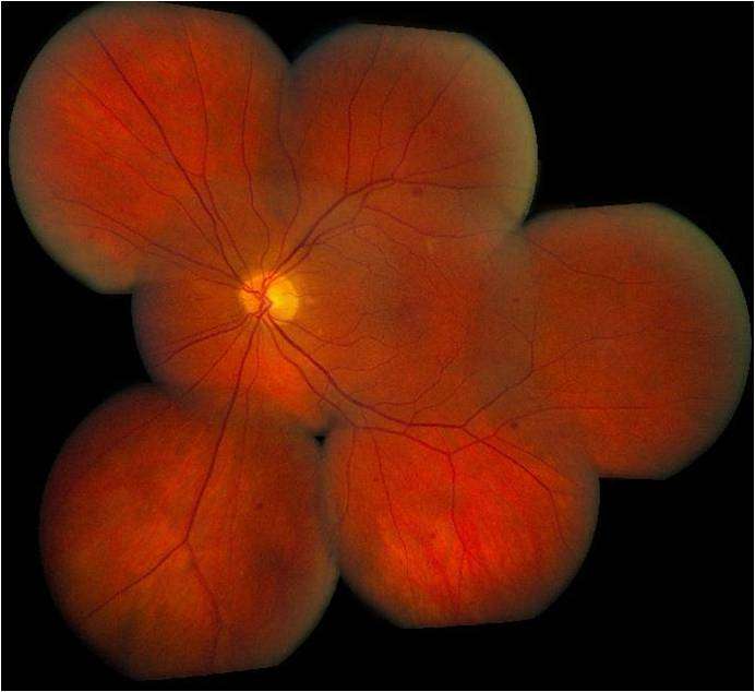
Retinopathy of prematurity (ROP) is a leading cause of blindness in infants. ROP (or Terry syndrome) is a disease of the eye affecting prematurely-born, low
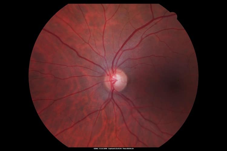
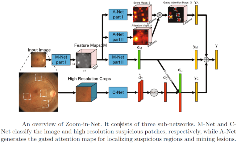
Image enhancement of retinal structures has the potential to facilitate diagnosis of several eye diseases. Retinal disease diagnosis and monitoring often requires very delicate analysis
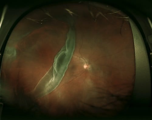
Retinal detachment occurs when part of the retina detaches itself from the pigmented cell layer of the RPE, depriving itself of blood and nutrition: this
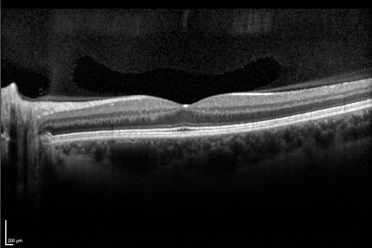
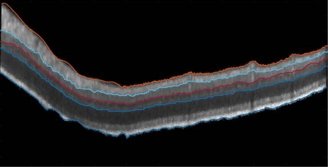
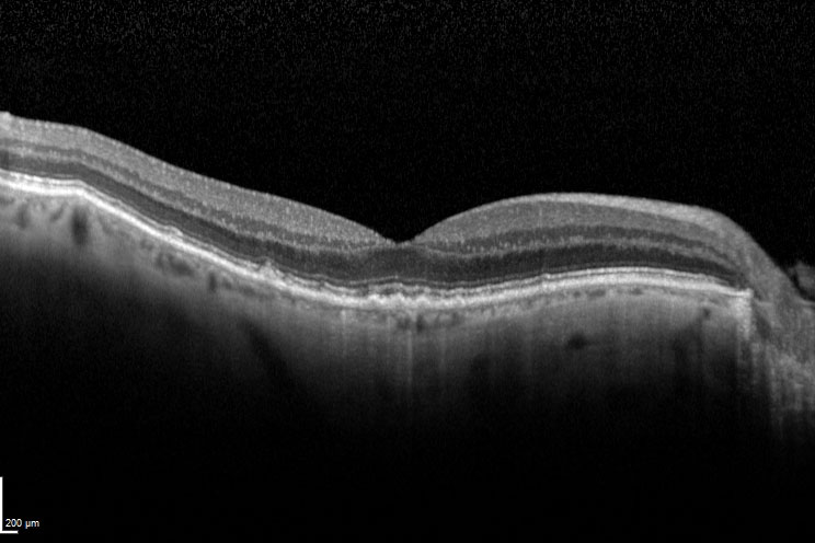
OCT is the only method that can perform noninvasive imaging with non-ionizing radiation and offering relatively good resolution. That is why it has become a
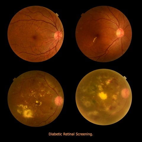
Diabetic Retinopathy (DR) is a leading cause of blindness, especially among adults and even more among the elderly segments of the population. It is associated
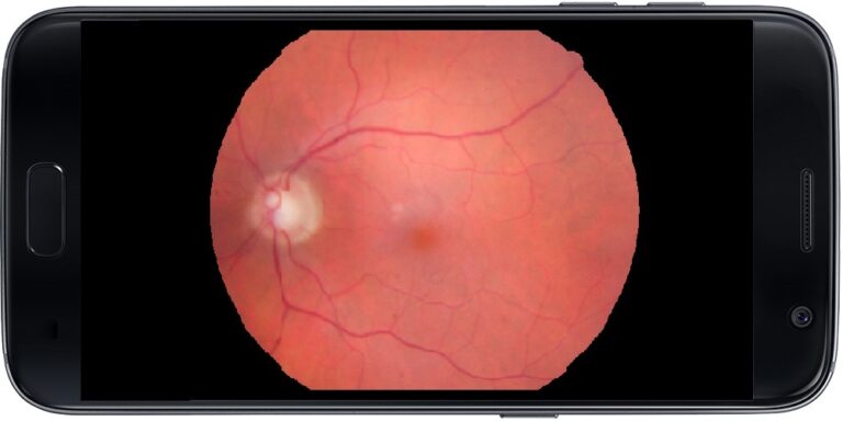
Portable cameras able to help ophthalmologists have been a desired solution for a long time. Among the reasons for the need of an additional device
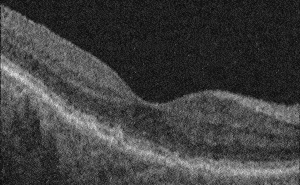
In a previous article, we talked about Geographic Atrophy segmentation in 2D images. This article focuses on how OCT images shed light on the development
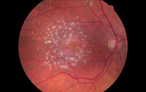
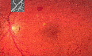
Geographic Atrophy (generally called GA) is a case of advanced Dry AMD (Age-related Macular Degeneration) which might lead to vision loss. As a consequence of