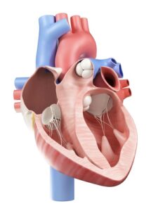
Left Ventricle Statistical Shape Modelling
Statistical shape modelling is a powerful tool for visualizing geometric and functional patterns of variation in all organs and also a reliable left ventricle shape model can prove itself very useful, though quite challenging to construct. RSIP Vision knows how to do this and many other image processing solutions for cardiology. And since cardiovascular diseases account for million of deaths per year in the developed world, this work is of vital importance.
Read More
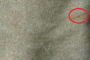
Fabric Inspection with Texture Analysis
Vision-based production inspection systems using camera-based scanning are now quite common in in-line production lines such as in steel, leather and fabrics manufacturing. Inspection is a crucial process since it can reduce process and enhance product quality. We recommend here a texture analysis for defect and novelty detection in fabrics and non-structured surfaces. Our fabric inspection algorithms are developed to detect deviations from local pattern and texture, anomalies and defects.

3D inspection and crack detection
Industrial production is prone to surface defects and it often needs to be inspected prior to shipment, when still in a semi-finished status. Cracks being very frequent in many types of material, vision-based crack inspection and detection is cost effective and offers high reproducibility and reliability. Here is a contact-free procedure using laser scanning, which can be placed in-line for continuous inspection during production.
Read More
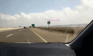
Road signs detection with machine learning
Frequent variations of speed limits (mainly due to roadwork and other maintenance), offer ADAS technology a chance to help drivers respect traffic laws and security. The traffic signs detection software developed by RSIP Vision detects a road sign at distance and verifies via machine learning if it is a speed limit sign and what that limit is. These detection and classification processes being based on machine learning, our application is able to do things that regular “engineered” software cannot do.
Read More
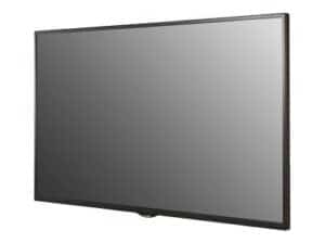
Flat Panel Display Inspection
This project compares the performance of a new inspection procedure of Flat Panel Displays (FPD) with the results obtained using a previously existing process. The goal was to demonstrate the correct detection and defect position reported by the new technology. This was done by putting in place a system in charge of image acquisition software and control which drives the captured frames to a sophisticated algorithmical registration analysis system developed by RSIP Vision.
Read More
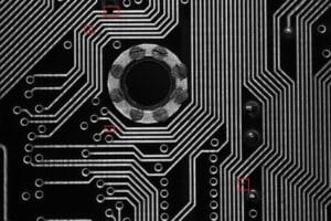
Wafer defect detection by feature matching
Detection of microscopic defect in wafers and printed circuit boards is a standard procedure in the manufacturing process. The time consuming human inspection has been replaced in nearly all production lines with an automatic in-line camera-based examination, which can be very effective usingcomputer vision and image processing technologies to detect any anomalies. Via algorithms of feature extraction and matching, RSIP Vision is able to track defects leading to dramatically improvements in reliability and usability.
Read More

Barcode Detection in complex environments
Automatically localizing and reading barcodes captured by a smartphone-based cameras have great value for industrial and personal applications but create new technical challenges: quality of the image, motion blurs, unpredictable distance to barcode, non-uniform orientation and unknown location of the barcode itself. Image processing and computer vision algorithms can solve these issues: ask RSIP Vision how we do it.
Read More

Real time OCR in natural scenes
With the advancement in technology, the demand for OCR in natural environment is growing, even though outdoor conditions are far from being optimal for machine vision applications: occlusion of written text, text orientation, font style, blurring due to camera motion, and lighting conditions can prove themselves significant challenges in the task of performing real time OCR. Great progress has recently been made in the recognition of characters partially occluded and under heavy noise. RSIP Vision tells you how.
Read More

Live cells tracking
Manual cell inspection is limited to tracking a relatively small number of cells in short periods of time and it is prone to human errors. On the other hand, computer vision algorithms can be used to perform fast scanning, segmentation and tracking of large cell populations over long periods of time. Taking advantage of our experience in segmentation, microscopy and machine learning, this procedure helps saving both labor and time, hence enabling more timely diagnostic and therapy.
Read More

Computer vision in Israel
The spectacular growth of digital imaging technology has made the solution to the problems of automated image interpretation much easier; the results are quite exciting
Airways segmentation with Deep Learning
Image processing is a fundamental technique in the quest to identify lung cancer, one of the main causes of death among both men and women, and many other lung pathologies. For the worst diseases, survival rate depends on the stage in which the disease is diagnosed and correct segmentation of airway vessels offers the most effective solution to determine the lesion’s size and location, significantly improving diagnosis and treatment. Our solution is built upon Deep Learning and neural networks.
Read More
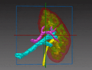
Kidney Segmentation
The most dramatically common kidney diseases are: kidney cancer, hitting 50,000 new patients every year only in the U.S.; and kidney failures, which leave the organ unable to remove wastes. Laparoscopic partial nephrectomy operations remove or reduce kidney tumors and some renal malfunctions. We at RSIP Vision help by providing a semi-automatic and very accurate kidney segmentation technique, built on deep learning and neural networks to create a kidney model which would be specific for each patient.
Read More
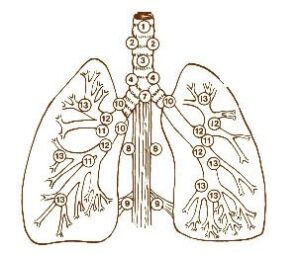
Pulmonary lobes segmentation
Emphysema quantification and lung nodule detection are among the clinical applications which benefit the most from lobes segmentation in CT scans. Proper lung segmentation is key to determine the boundaries of lobes and prevent pleural damage during examination and treatment. When correctly located, diseases are treated faster and better, hence the call for RSIP Vision to find a faster alternative to time-consuming manual segmentation.
Read More
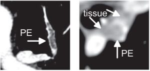
Pulmonary embolism detection
Timely detection of pulmonary embolism via CT angiography is key to reduce mortality risks. Human detection of pulmonary embolism (being often too slow, RSIP Vision recommends a combination of bidimensional and tridimensional image-processing techniques to achieve computer-aided pulmonary embolism detection which enables prompter diagnosis and treatment.
Read More

Date sorting
RSIP Vision has successfully worked in number of dates grading (or dates sorting) projects for our clients. Our automatic fruit recognition system is able to identify with high speed and accuracy all meaningful product features such as size, weight, defect, quality, color, texture, ripeness and others, offering key benefits to our clients: namely, fast and high-volume classification, savings in labor costs, consistent quality and reduced time-to-market.
Read More
Lung Lymph Nodes Detection
Analyzing pulmonary lymph nodes can give us valuable information for lung cancer diagnosis and treatment. This solution too uses advanced algorithm of computer vision for pulmonology; it also allows to overcome technical difficulties like low image contrast and high nodes variation, offering a drastic improvement over techniques currently used to detect lung lymph nodes.
Read More
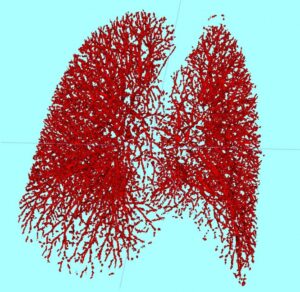
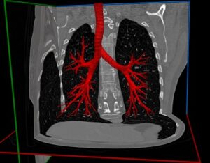
Lung vessel segmentation
Blood vessel segmentation of the lungs can help to identify important pulmonary diseases, characterizing nodules in the lungs, detecting pulmonary emboli and evaluating the lungs vasculature. Our technique of automatic pulmonary vessel segmentation completes very effectively the vessel tree structure provided by the CT scans of the lung, in such a way that the resulting image is more precise and matchlessly faster than any manual segmentation could be.
Read More
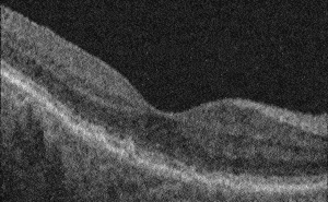
Measurement of Retinal Thickness
Retinal thickness is a key measurement used to assess the health of the retina and whether it needs any treatment. Thickness measures can be compared to optimal ranges or to data from the same patient over time, helping ophthalmologists to identify retinal disorders. Advanced optimization methods, borrowed from graph theory, enable us to solve the complex challenge of measuring retinal thickness within reasonable processing time.
Read More
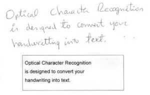
Deep Learning For OCR
OCR used for the visual inspection of documents has found wide application in both industry and research, though it is more commonly found in connection with printed characters than for handwritten ones, mainly owing to the variability in handwritten characters’ shapes and styles. Hence the need for automatic recognition performed by vision-based tailor-made algorithms and adjustment. Deep Neural Networks as a learning mechanism to perform recognition have proved to be particularly powerful tools, due to their high accuracy in both spotting text region and deciphering the characters.
Read More
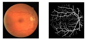
Vessel Segmentation Using Deep Learning
Various segmentation methods, whether based on Convolution Neural Networks or traditional image processing techniques, can be used to delineate the vascular tree in clinical imaging. Given the few features distinguishing veins from arteries (usually brighter and thinner than veins), the challenge consists of training a binary classifier assigning each pixel to the category of vein or artery. This article covers the advantages of using CNNs and deep neural networks for the classification and segmentation of vessels in fundus images.
Read More