Skip to content
  • Our Work
    • Fields
      • Cardiology
      • ENT
      • Gastro
      • Orthopedics
      • Ophthalmology
      • Pulmonology
      • Surgical
      • Urology
      • Other
    • Modalities
      • Endoscopy
      • Medical Segmentation
      • Microscopy
      • Ultrasound
  • Success Stories
  • Insights
    • Magazine
    • Upcoming Events
    • Webinars
    • Meetups
    • News
    • Blog
  • The company
    • About us
    • Careers
Menu
  • Our Work
    • Fields
      • Cardiology
      • ENT
      • Gastro
      • Orthopedics
      • Ophthalmology
      • Pulmonology
      • Surgical
      • Urology
      • Other
    • Modalities
      • Endoscopy
      • Medical Segmentation
      • Microscopy
      • Ultrasound
  • Success Stories
  • Insights
    • Magazine
    • Upcoming Events
    • Webinars
    • Meetups
    • News
    • Blog
  • The company
    • About us
    • Careers
Contact

Lungs tumors and nodules segmentation with Deep Learning

Lung nodules are masses of tissue in the lung which on a chest X-ray or computerized tomography (CT) scan appear as round, white shadows. Though lung nodules are quite common and most of them are noncancerous (benign), some nodules may be cancerous and it is extremely important to identify them early in their course, when they can still be effectively treated and cured. In addition, segmentation of lung tumors helps finding out how far they have spread (staging), their response to treatments as well as their progression in size and shape.
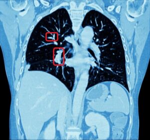
Medical experts can visually identify nodules on a CT scan, but they need to rely on computerized analysis to estimate shape and volume of nodules, as well as their progression over time. It is visually easier to identify nodules than lung tumors, since the former are supposed to have an elliptical shape, while the chromatic aspect of the latter is difficult to distinguish from healthy tissues on a CT image. This difference reflects also on the annotation phase: it will be easier for a non-expert annotator to identify a tumor than a nodule.

Lung nodule segmentation
Slice of segmentation result of a nodule from CT scan

Automatic lung nodules segmentation

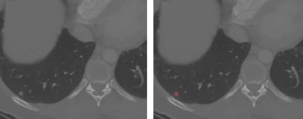
Our client needed an automatic process of lung nodules and tumors segmentation to deal with this difficulty in a way that is quick to perform and memory efficient. Our methodology (once built around graph cuts algorithms) currently focuses on Deep Learning with neural networks. Their advantages are multiple: besides offering a much faster segmentation, this is a fully automated solution, able to automatically detect all tumors and nodules, with no need for external intervention. This solution proves to provide a simple and effective answer to many problems of medical segmentation. It is robust and practical, as it solves the challenges fast and efficiently. We at RSIP Vision (and not only us) consider it the best approach to perform nodule and tumor detection and segmentation in lung CT images.
Lung tumor

Lung tumor - zoom
Detection and segmentation of a tumor in a CT scan

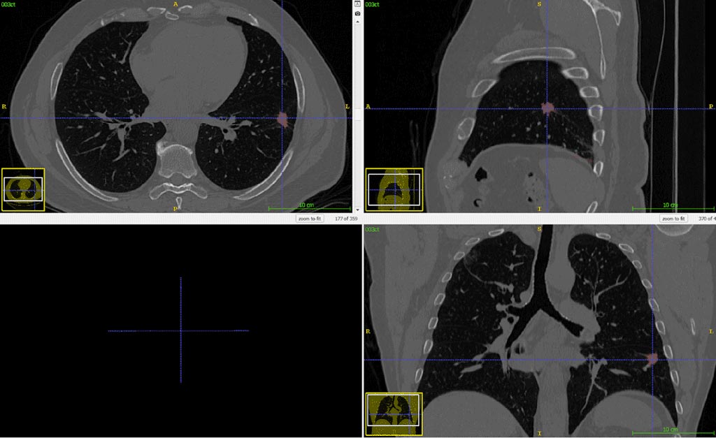
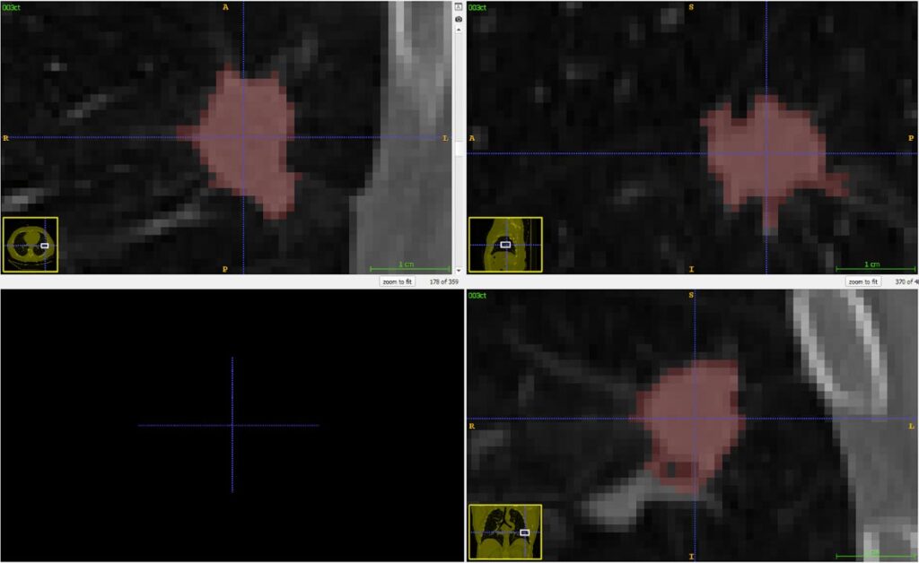
Compared with hand-made nodule segmentation in the lung (performed by an expert), this automatic lung nodules and tumors segmentation method grants results which are very close to the (much slower) manual segmentation. It also enables our client to overcome the difficulty of tumors and healthy tissues appearing on the CT scan in a very similar range of intensity values. You can read here about other projects led by RSIP Vision in the field of pulmonology.

Share

Share on linkedin
Share on twitter
Share on facebook

Main Field

Pulmonology

RSIP Vision’s image processing expertise in medical imaging is currently used in numerous projects, including many applications of computer vision in pulmonology. We are very proud of our contribution to lung medicine: feature detection, lung segmentation and countless other works in the pulmonary imaging field enabled our clients to give the means to physicians to save lives by giving faster and more appropriate treatment to all kind of lung diseases. You can read below about some of our breakthrough developments in Pulmonology and Bronchoscopy.

View Pulmonology

Categories

  • Pulmonology, RSIP Vision Learns

Related Content

Pulmonary embolism

Detecting Pulmonary Embolism from CT Scan

Lung vasculature segmentation

Lung tumors

Lung Tumor Segmentation

Lung Nodules Segmentation

Lung fissures

Lung Fissures Segmentation

Visible lung cancer on CT scan of chest and abdomen

Chest CT Scan Analysis with Deep Learning

Pulmonary embolism

Detecting Pulmonary Embolism from CT Scan

Lung vasculature segmentation

Lung tumors

Lung Tumor Segmentation

Lung Nodules Segmentation

Lung fissures

Lung Fissures Segmentation

Visible lung cancer on CT scan of chest and abdomen

Chest CT Scan Analysis with Deep Learning

Show all

RSIP Vision

Field-tested software solutions and custom R&D, to power your next medical products with innovative AI and image analysis capabilities.

Read more about us

Get in touch

Please fill the following form and our experts will be happy to reply to you soon

Recent News

IBD Scoring – Clario, GI Reviewers and RSIP Vision Team Up

RSIP Neph Announces a Revolutionary Intra-op Solution for Partial Nephrectomy Surgeries

Announcement – RSIP Vision Presents Successful Preliminary Results from Clinical Study of 2D-to-3D Knee Bones Reconstruction

Announcement – New Urological AI Tool for 3D Reconstruction of the Ureter

All news
Upcoming Events
Stay informed for our next events
Subscribe to Our Magazines

Subscribe now and receive the Computer Vision News Magazine every month to your mailbox

 
Subscribe for free
Follow us
Linkedin Twitter Facebook Youtube

contact@rsipvision.com

Terms of Use

Privacy Policy

© All rights reserved to RSIP Vision 2023

Created by Shmulik

  • Our Work
    • title-1
      • Ophthalmology
      • Uncategorized
      • Ophthalmology
      • Pulmonology
      • Cardiology
      • Orthopedics
    • Title-2
      • Orthopedics
  • Success Stories
  • Insights
  • The company