Skip to content
  • Our Work
    • Fields
      • Cardiology
      • ENT
      • Gastro
      • Orthopedics
      • Ophthalmology
      • Pulmonology
      • Surgical
      • Urology
      • Other
    • Modalities
      • Endoscopy
      • Medical Segmentation
      • Microscopy
      • Ultrasound
  • Success Stories
  • Insights
    • Magazine
    • Upcoming Events
    • Webinars
    • Meetups
    • News
    • Blog
  • The company
    • About us
    • Careers
Menu
  • Our Work
    • Fields
      • Cardiology
      • ENT
      • Gastro
      • Orthopedics
      • Ophthalmology
      • Pulmonology
      • Surgical
      • Urology
      • Other
    • Modalities
      • Endoscopy
      • Medical Segmentation
      • Microscopy
      • Ultrasound
  • Success Stories
  • Insights
    • Magazine
    • Upcoming Events
    • Webinars
    • Meetups
    • News
    • Blog
  • The company
    • About us
    • Careers
Contact

Chromosome classification

Chromosomes are packaged and organized structures which contain most of the DNA of living organisms. The DNA that constitutes the chromosomes becomes more tightly aligned in the middle of the cell during the metaphase (the cell division) and is then visible on a microscope.
In humans, each cell generally contains 23 pairs of chromosomes (totaling 46 chromosomes): 22 pairs are called autosomes and look the same in females and males. The 23rd pair (the two sex chromosomes) differ between females and males in that females have two X chromosomes and males have one X and one Y chromosome.

Karyotype

The karyotype test is done to identify and evaluate size, shape, and number of chromosomes in the body cells. Additional or missing chromosomes, as well as abnormal location of chromosome pieces, can trigger major troubles to an individual’s growth, development and body functioning.
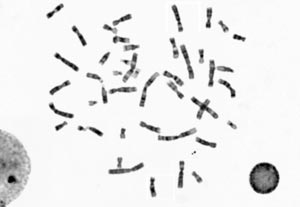
Several features help identify the chromosome within the individual karyotype set: the distribution of gray shades along the medial axis, the position of the centromere (a limited region of the chromosome, which might be challenging to identify) and the relative length of the chromosome in the set.
Cytologists able to perform a manual chromosome classification are rare and the results they obtain could be improved in terms of time, and costs when supported or replaced by machine learning systems. Most of the research activity in computer vision in recent years show very high capabilities on different types of datasets and applications and RSIP Vision has developed much experience in the machine learning field.

Chromosomes classification with deep learning

We decided to develop a solution to our client’s need by means of convolutional neural networks (CNN), a deep learning technique currently deployed in many practical applications like image recognition, speech recognition, photo taggers and many more. CNN are extremely valuable when nearby values of array data are correlated. Ideally, the convolutional neural networks should be trained on a very large dataset. It can also be trained on one system with sufficient number of cases and then fine-tuned to work on a new system.
We used the convolutional neural networks in first place as feature extractor; then we aligned and centered the data; finally, we extracted the features map from each image and trained them on a linear 1d classifier.
Our client received a very robust solution for chromosome classification, which will keep becoming more and more solid as it is given additional datasets to train on.
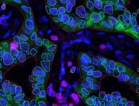
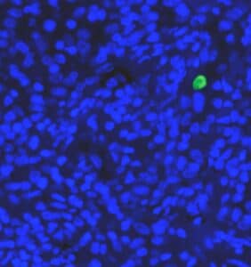
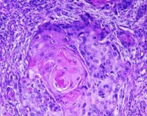
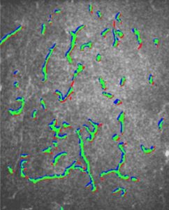
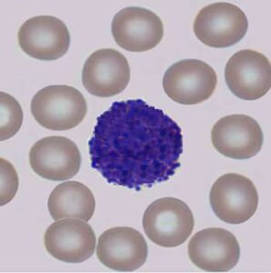
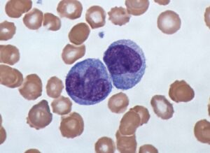
 

Share

Share on linkedin
Share on twitter
Share on facebook

Main Field

Microscopy

View Microscopy

Categories

  • Microscopy, RSIP Vision Learns

Related Content

Multiplex

Multiplex IF Analysis

CTCs - Circulating Tumor Cells

Circulating Tumor Cells (CTCs)

Pharma - Tissue Analysis

Tissue Analysis with AI

Pharma - Dendritic cell

Detection and Segmentation of Dendritic Cells

Cell Classification - pattern recognition classification techniques

Biomedical image processing and algorithms

Microscopy view of Monocytes

Cell Classification software

Show all

RSIP Vision

Field-tested software solutions and custom R&D, to power your next medical products with innovative AI and image analysis capabilities.

Read more about us

Get in touch

Please fill the following form and our experts will be happy to reply to you soon

Recent News

IBD Scoring – Clario, GI Reviewers and RSIP Vision Team Up

RSIP Neph Announces a Revolutionary Intra-op Solution for Partial Nephrectomy Surgeries

Announcement – RSIP Vision Presents Successful Preliminary Results from Clinical Study of 2D-to-3D Knee Bones Reconstruction

Announcement – New Urological AI Tool for 3D Reconstruction of the Ureter

All news
Upcoming Events
Stay informed for our next events
Subscribe to Our Magazines

Subscribe now and receive the Computer Vision News Magazine every month to your mailbox

 
Subscribe for free
Follow us
Linkedin Twitter Facebook Youtube

contact@rsipvision.com

Terms of Use

Privacy Policy

© All rights reserved to RSIP Vision 2023

Created by Shmulik

  • Our Work
    • title-1
      • Ophthalmology
      • Uncategorized
      • Ophthalmology
      • Pulmonology
      • Cardiology
      • Orthopedics
    • Title-2
      • Orthopedics
  • Success Stories
  • Insights
  • The company