Skip to content
  • Our Work
    • Fields
      • Cardiology
      • ENT
      • Gastro
      • Orthopedics
      • Ophthalmology
      • Pulmonology
      • Surgical
      • Urology
      • Other
    • Modalities
      • Endoscopy
      • Medical Segmentation
      • Microscopy
      • Ultrasound
  • Success Stories
  • Insights
    • Magazine
    • Upcoming Events
    • Webinars
    • Meetups
    • News
    • Blog
  • The company
    • About us
    • Careers
Menu
  • Our Work
    • Fields
      • Cardiology
      • ENT
      • Gastro
      • Orthopedics
      • Ophthalmology
      • Pulmonology
      • Surgical
      • Urology
      • Other
    • Modalities
      • Endoscopy
      • Medical Segmentation
      • Microscopy
      • Ultrasound
  • Success Stories
  • Insights
    • Magazine
    • Upcoming Events
    • Webinars
    • Meetups
    • News
    • Blog
  • The company
    • About us
    • Careers
Contact

Kidney Segmentation

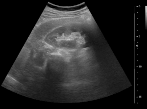
Healthy kidneys have an essential part of proper bodily function. Therefore, early diagnosis and treatment of kidney pathologies is crucial. As people age, some will suffer from a variety of kidney pathologies, most common are kidney stones (nephrolithiasis), but some people may suffer from kidney tumors. Unfortunately, some of these pathologies can greatly affect the quality of life, and some tumors are cancerous. Therefore, many patients need further assessment and treatment. In some cases, contrast enhanced computed tomography (CECT) is indicated for renal imaging. CECT allows the radiologist to evaluate the kidney cortex, medulla, calyces, ureter and vessels. Therefore, kidney segmentation from abdominal computed tomography (CT) images is a prerequisite step towards automated computerized diagnosis of renal pathologies, and treatment of kidney stones including ureteroscopy and percutaneous nephrolithotomy (PCNL). In addition, it is a useful tool for surgical planning of kidney tumors resection and ablation.

Kidney segmentation
Pink = Renal artery; Light blue = Renal vein; Yellow = Renal calyces and ureter; Red/Maroon = Renal cortex; Green = Renal medulla

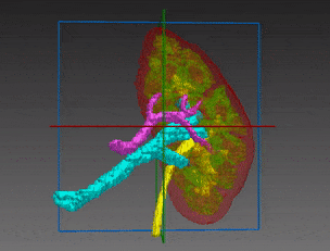
The complexity of kidney segmentation stems from similar density levels of adjacent structures, e.g the liver and spleen and high variability of shape and anatomical position of the kidneys. Number of calyces varies and they have no known tree structure, with a large interpatient variation.

Automated kidney segmentation

RSIP Vision has developed an impressive image processing algorithm for abdominal CT scans that segments the kidney region with high accuracy. This kidney segmentation is also the first step which may enable automatic detection of renal pathologies in the next step.
CT urography in the examination of choice for detecting renal and collecting system pathologies. It consists of four stages – unenhanced, corticomedullary/arterial, nephrographic/parenchymal and urographic phases. Each phase emphasizes different parts of the renal system. The unenhanced phase is used for identifying calcified structures such as stones, corticomedullary allows to detect renal arteries and renal cell carcinoma (RCC), nephrographic phase helps with identifying veins and differentiation between simple cysts and other masses and the urographic phase enhances the collecting system, revealing any malformations or urothelial tumors such as transitional cell carcinoma (TCC).
Since kidneys are a relatively small organ, all the tiny details within them must be accurately annotated. In the same line, kidney blood-vessels should be segmented with care. RSIP Vision’s kidney segmentation module is the ideal tool to be used prior to kidney surgical procedures.
A U-Net based neural network was utilized for this task, enabling the learning algorithm to pick up on various important features from within the scan and out-perform any other technique we used, yielding great quality segmentations. Do you have medical segmentation projects? Call us!

Share

Share on linkedin
Share on twitter
Share on facebook

Main Field

Urology

Urology is one of the many medical fields that are being revolutionized by Artificial Intelligence. Prostate, bladder, urethra, kidneys and more key organs are already benefitting from impressive breakthroughs brought about by new technologies. AI is now able to contribute to more accurate and faster detection, segmentation, classification and diagnose for many urology disfunctions and diseases, including cancer, BPH and urolithiasis. Thanks to the latest generation of AI and computer vision techniques (deep learning in particular), AI provides now a precious support to the medical team in the operating room as well as during all stages of treatment, drastically improving the quality of therapy and reducing recovery time. You can find below some of RSIP Vision's projects and pioneering research in the field of AI for urology.

View Urology

Categories

  • RSIP Vision Learns, Urology

Related Content

Neph - Partial Nephrectomy surgery

RSIP Neph Announces a Revolutionary Intra-op Solution for Partial Nephrectomy Surgeries

Benign Prostatic Hyperplasia BPH

AI for Benign Prostatic Hyperplasia BPH

Stone tracking - urology

Stone tracking during kidney stone removal

Tissue Sparing in urology

AI-Assisted Tissue Sparing in Urology

Percutaneous Nephrolithotomy

PCNL – Planning and real-time navigation

Prostate Tumor Segmentation

Implementing AI to Improve PI-RADS Scoring

Show all

RSIP Vision

Field-tested software solutions and custom R&D, to power your next medical products with innovative AI and image analysis capabilities.

Read more about us

Get in touch

Please fill the following form and our experts will be happy to reply to you soon

Recent News

IBD Scoring – Clario, GI Reviewers and RSIP Vision Team Up

RSIP Neph Announces a Revolutionary Intra-op Solution for Partial Nephrectomy Surgeries

Announcement – RSIP Vision Presents Successful Preliminary Results from Clinical Study of 2D-to-3D Knee Bones Reconstruction

Announcement – New Urological AI Tool for 3D Reconstruction of the Ureter

All news
Upcoming Events
Stay informed for our next events
Subscribe to Our Magazines

Subscribe now and receive the Computer Vision News Magazine every month to your mailbox

 
Subscribe for free
Follow us
Linkedin Twitter Facebook Youtube

contact@rsipvision.com

Terms of Use

Privacy Policy

© All rights reserved to RSIP Vision 2023

Created by Shmulik

  • Our Work
    • title-1
      • Ophthalmology
      • Uncategorized
      • Ophthalmology
      • Pulmonology
      • Cardiology
      • Orthopedics
    • Title-2
      • Orthopedics
  • Success Stories
  • Insights
  • The company