Skip to content
  • Our Work
    • Fields
      • Cardiology
      • ENT
      • Gastro
      • Orthopedics
      • Ophthalmology
      • Pulmonology
      • Surgical
      • Urology
      • Other
    • Modalities
      • Endoscopy
      • Medical Segmentation
      • Microscopy
      • Ultrasound
  • Success Stories
  • Insights
    • Magazine
    • Upcoming Events
    • Webinars
    • Meetups
    • News
    • Blog
  • The company
    • About us
    • Careers
Menu
  • Our Work
    • Fields
      • Cardiology
      • ENT
      • Gastro
      • Orthopedics
      • Ophthalmology
      • Pulmonology
      • Surgical
      • Urology
      • Other
    • Modalities
      • Endoscopy
      • Medical Segmentation
      • Microscopy
      • Ultrasound
  • Success Stories
  • Insights
    • Magazine
    • Upcoming Events
    • Webinars
    • Meetups
    • News
    • Blog
  • The company
    • About us
    • Careers
Contact

Prostate segmentation in MR images

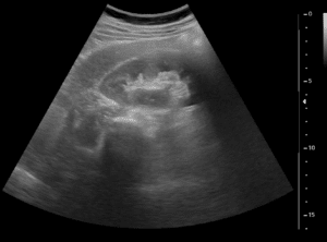
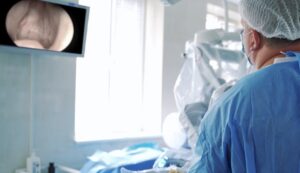
The prostate gland is part of the male reproductive system. It is found in front of the rectum and below the bladder, surrounding the first part of the urethra. Ultrasound and MRI are the most commonly used techniques to image the prostate gland. In particular, detailed MR images allow to evaluate the prostate and determine the presence of diseases.

The primary indication for MRI of the prostate is the evaluation of prostate cancer. The test is commonly used to evaluate the extent of prostate cancer in order to determine if it is confined to the prostate gland, or if it has spread outside. The images can then be examined by an expert physician to evaluate and decide the most appropriate treatment response.

Volume is a key indicator of the health of the prostate: it reveals key information about the stage of the cancer, hints at the probable prognosis and suggests possible treatments. Besides the dangers of underestimated or late detection of an aggressive fast-growing tumor, it is crucial to avoid also overdiagnosis and overtreatment of a slow tumor, which is unlikely to threaten a man’s life.

The need for reliable evidence is far from trivial. Prostate cancer is the second most common cancer among American men. More than 200,000 new cases of prostate cancer are diagnosed every year and about 1 man in 7 will be diagnosed with prostate cancer during his lifetime.

Prostate Segmentation

Precise estimate of the prostate volume

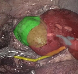
It is therefore key to get a most precise estimate of the prostate volume. When the determination of this information is inaccurate and/or inconsistent, serious limitations are posed to its usefulness in clinical practice: size, shape, and location of the prostate in relation to adjacent organs is also essential information while planning surgeries (like prostatectomy), radiation therapy, and new minimally invasive therapies, such as cryotherapy and high-intensity focused ultrasound.

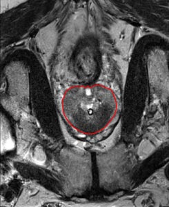
MRI-guided prostate volume estimation

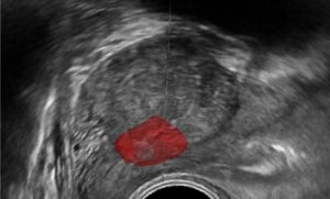
The rich experience of RSIP Vision in medical segmentation (read here about kidney segmentation) enables us to recommend an approach based on a semi-automatic prostate segmentation, in which the initial input is manual (coming from the physician). The method follows an optimal path finding technique, a robotic system able to deal with image noise. The optimal path technique starts off from ultrasound imaging, which contains noise-rich data particularly complex to analyze. The technique finds the borders of the prostate, enabling a reliable calculation of the prostate volume, leading to the key information which the health professional is looking for. This approach is giving our client and us very robust, high quality results which are proving extremely helpful in clinical practice for both detection and diagnostic.

Share

Share on linkedin
Share on twitter
Share on facebook

Main Field

Urology

Urology is one of the many medical fields that are being revolutionized by Artificial Intelligence. Prostate, bladder, urethra, kidneys and more key organs are already benefitting from impressive breakthroughs brought about by new technologies. AI is now able to contribute to more accurate and faster detection, segmentation, classification and diagnose for many urology disfunctions and diseases, including cancer, BPH and urolithiasis. Thanks to the latest generation of AI and computer vision techniques (deep learning in particular), AI provides now a precious support to the medical team in the operating room as well as during all stages of treatment, drastically improving the quality of therapy and reducing recovery time. You can find below some of RSIP Vision's projects and pioneering research in the field of AI for urology.

View Urology

Categories

  • RSIP Vision Learns, Urology

Related Content

Neph - Partial Nephrectomy surgery

RSIP Neph Announces a Revolutionary Intra-op Solution for Partial Nephrectomy Surgeries

Benign Prostatic Hyperplasia BPH

AI for Benign Prostatic Hyperplasia BPH

Stone tracking - urology

Stone tracking during kidney stone removal

Tissue Sparing in urology

AI-Assisted Tissue Sparing in Urology

Percutaneous Nephrolithotomy

PCNL – Planning and real-time navigation

Prostate Tumor Segmentation

Implementing AI to Improve PI-RADS Scoring

Show all

RSIP Vision

Field-tested software solutions and custom R&D, to power your next medical products with innovative AI and image analysis capabilities.

Read more about us

Get in touch

Please fill the following form and our experts will be happy to reply to you soon

Recent News

IBD Scoring – Clario, GI Reviewers and RSIP Vision Team Up

RSIP Neph Announces a Revolutionary Intra-op Solution for Partial Nephrectomy Surgeries

Announcement – RSIP Vision Presents Successful Preliminary Results from Clinical Study of 2D-to-3D Knee Bones Reconstruction

Announcement – New Urological AI Tool for 3D Reconstruction of the Ureter

All news
Upcoming Events
Stay informed for our next events
Subscribe to Our Magazines

Subscribe now and receive the Computer Vision News Magazine every month to your mailbox

 
Subscribe for free
Follow us
Linkedin Twitter Facebook Youtube

contact@rsipvision.com

Terms of Use

Privacy Policy

© All rights reserved to RSIP Vision 2023

Created by Shmulik

  • Our Work
    • title-1
      • Ophthalmology
      • Uncategorized
      • Ophthalmology
      • Pulmonology
      • Cardiology
      • Orthopedics
    • Title-2
      • Orthopedics
  • Success Stories
  • Insights
  • The company