Skip to content
  • Our Work
    • Fields
      • Cardiology
      • ENT
      • Gastro
      • Orthopedics
      • Ophthalmology
      • Pulmonology
      • Surgical
      • Urology
      • Other
    • Modalities
      • Endoscopy
      • Medical Segmentation
      • Microscopy
      • Ultrasound
  • Success Stories
  • Insights
    • Magazine
    • Upcoming Events
    • Webinars
    • Meetups
    • News
    • Blog
  • The company
    • About us
    • Careers
Menu
  • Our Work
    • Fields
      • Cardiology
      • ENT
      • Gastro
      • Orthopedics
      • Ophthalmology
      • Pulmonology
      • Surgical
      • Urology
      • Other
    • Modalities
      • Endoscopy
      • Medical Segmentation
      • Microscopy
      • Ultrasound
  • Success Stories
  • Insights
    • Magazine
    • Upcoming Events
    • Webinars
    • Meetups
    • News
    • Blog
  • The company
    • About us
    • Careers
Contact

Pulmonary lobes segmentation

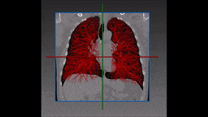
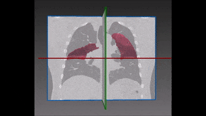
The left lung is divided in two lobes, the upper and the lower lobe. These two lobes are separated by an oblique fissure (the left major fissure). In the right lung, an oblique fissure (the right minor fissure) and a horizontal fissure (the right major fissure) separate the lung into three lobes – the upper, the middle, and the lower lobe. Each lobe has its own pleural covering. The two human lungs are thus divided in five lobes.

Lung lobes segmentation is particularly important during the process of assessing the location and progression of diseases, as well as when choosing their most appropriate treatment. Emphysema quantification and lung nodule detection are among the clinical applications which benefit the most from lung segmentation in CT scans. In fact, proper lung segmentation is key to determine the boundaries of lobes and prevent pleural damage during examination and treatment. When correctly located, diseases can be treated faster and better, hence the need to find a faster alternative to manual segmentation, which is time consuming and therefore far from ideal.

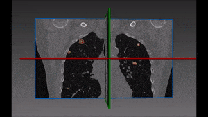
The main obstacle to lung segmentation comes from differences in anatomy and deformed or incomplete fissures: in fact, anatomical variations are the reason why atlas registration gives better results when performed on healthy and regular lungs. The results of atlas and multi-atlas approaches are less satisfactory when the lungs differ significantly from the atlas because of ailments, shape variations or simply an incomplete fissure on the CT scan.

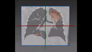
Our client needed a solution to perform fast and reliable pulmonary lobes segmentations. Among the many approaches which have been recently tested by the scientific community, we achieve the best result by combining several techniques around a watershed transformation.

Our approach at RSIP Vision starts with a segmentation of the pulmonary vessels: this phase is particularly helpful since it is assumed that no major blood vessel is to be found in the proximity of the fissures. This first step enables to separate the pulmonary vasculature from other dense structures which are more suitable to be fissures candidates. This is done using information coming from vessels, fissures and bronchi while giving a fissure probability to each voxel by the way of a Hessian matrix fissure enhancement procedure.

The enhancement of the fissures which is implemented with this technique attributes to each voxel a fissure similarity value comprised between 0 and 1. These results are then converted to a segmentation of the lobe fissures, following criteria such as minimal similarity and range of intensity.

The following step of our solution is built on our own variation of a watershed transformation technique performed starting from an initially segmented fissure and successively extended to the surrounding region of interest. The information coming from vessels, fissures and bronchi is thus combined into a cost image which, together with 3D markers of the five lobes, is used as input of this watershed transformation.

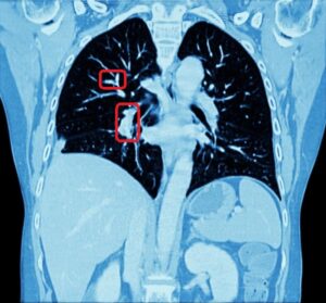
The robustness of this lobe segmentation has been successfully tested on a set of lungs showing incomplete fissures, since these are the most challenging cases in lung segmentation. The results of the comparison prove that a correct segmentation can show far better results than those which are immediately visible on a CT image.

Share

Share on linkedin
Share on twitter
Share on facebook

Main Field

Pulmonology

RSIP Vision’s image processing expertise in medical imaging is currently used in numerous projects, including many applications of computer vision in pulmonology. We are very proud of our contribution to lung medicine: feature detection, lung segmentation and countless other works in the pulmonary imaging field enabled our clients to give the means to physicians to save lives by giving faster and more appropriate treatment to all kind of lung diseases. You can read below about some of our breakthrough developments in Pulmonology and Bronchoscopy.

View Pulmonology

Categories

  • Pulmonology, RSIP Vision Learns

Related Content

Pulmonary embolism

Detecting Pulmonary Embolism from CT Scan

Lung vasculature segmentation

Lung tumors

Lung Tumor Segmentation

Lung Nodules Segmentation

Lung fissures

Lung Fissures Segmentation

Visible lung cancer on CT scan of chest and abdomen

Chest CT Scan Analysis with Deep Learning

Pulmonary embolism

Detecting Pulmonary Embolism from CT Scan

Lung vasculature segmentation

Lung tumors

Lung Tumor Segmentation

Lung Nodules Segmentation

Lung fissures

Lung Fissures Segmentation

Visible lung cancer on CT scan of chest and abdomen

Chest CT Scan Analysis with Deep Learning

Show all

RSIP Vision

Field-tested software solutions and custom R&D, to power your next medical products with innovative AI and image analysis capabilities.

Read more about us

Get in touch

Please fill the following form and our experts will be happy to reply to you soon

Recent News

IBD Scoring – Clario, GI Reviewers and RSIP Vision Team Up

RSIP Neph Announces a Revolutionary Intra-op Solution for Partial Nephrectomy Surgeries

Announcement – RSIP Vision Presents Successful Preliminary Results from Clinical Study of 2D-to-3D Knee Bones Reconstruction

Announcement – New Urological AI Tool for 3D Reconstruction of the Ureter

All news
Upcoming Events
Stay informed for our next events
Subscribe to Our Magazines

Subscribe now and receive the Computer Vision News Magazine every month to your mailbox

 
Subscribe for free
Follow us
Linkedin Twitter Facebook Youtube

contact@rsipvision.com

Terms of Use

Privacy Policy

© All rights reserved to RSIP Vision 2023

Created by Shmulik

  • Our Work
    • title-1
      • Ophthalmology
      • Uncategorized
      • Ophthalmology
      • Pulmonology
      • Cardiology
      • Orthopedics
    • Title-2
      • Orthopedics
  • Success Stories
  • Insights
  • The company