Skip to content
  • Our Work
    • Fields
      • Cardiology
      • ENT
      • Gastro
      • Orthopedics
      • Ophthalmology
      • Pulmonology
      • Surgical
      • Urology
      • Other
    • Modalities
      • Endoscopy
      • Medical Segmentation
      • Microscopy
      • Ultrasound
  • Success Stories
  • Insights
    • Magazine
    • Upcoming Events
    • Webinars
    • Meetups
    • News
    • Blog
  • The company
    • About us
    • Careers
Menu
  • Our Work
    • Fields
      • Cardiology
      • ENT
      • Gastro
      • Orthopedics
      • Ophthalmology
      • Pulmonology
      • Surgical
      • Urology
      • Other
    • Modalities
      • Endoscopy
      • Medical Segmentation
      • Microscopy
      • Ultrasound
  • Success Stories
  • Insights
    • Magazine
    • Upcoming Events
    • Webinars
    • Meetups
    • News
    • Blog
  • The company
    • About us
    • Careers
Contact

Automatic segmentation of tumor cells

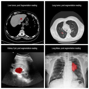
Molecular analysis of in histology is a powerful tool to explore prognostic markers after tissue staining. High throughput analysis of histological tissue enables quantification of abnormality in a given tissue, assess patient condition, and devise treatment. Tissue samples taken in biopsy allow researchers to screen for therapeutic agents by exposing the sample to a combination of drugs. Both expression levels and tissue morphological changes indicate response to a certain combination of drugs and can orient therapeutic avenue.

Manual segmentation of tumor cells:

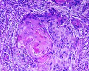
Common practice in histological analysis is to have two or more experts score the tissue response pattern. This visual examination is highly time-consuming and not readily available in clinical applications, where rapid intervention is crucial. Even for research purposes, tissue sampled in biopsy might not accurately capture the bulk tumor due to its irregular non-cylindrical shape. Therefore, segmentation of tumor cells by humans is quite unpractical and estimating the tumor burden becomes a non-trivial task even to human experts.

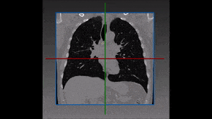
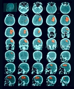
Fast scanning and analysis of histological tissue can be achieved by employing computer vision algorithm. These algorithms can autonomously perform quantification tasks, like estimating subpopulation fraction. Alternatively, they can achieve a semi-automated segmentation of tumor cells, working cooperatively with the human expert.

Squamous cell carcinoma

Automated tumor cells segmentation:

Quantification of biomarker expression in a cancer sample tissue is based on segmentation and classification of the image into several classes. At first, tissue stains need to be de-convolved to examine the expression distribution among cells belonging to the tissue under observation. The color histograms are basic tools allowing separation of the image into several independent color channel histograms, allowing grouping of pixels into classes.

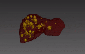
Next, morphological analysis of the cells forms a second level of separation. Abnormalities in cells often are detected by nuclear shape abnormalities. Segmentation of nuclei can be performed on the image histogram by maximal entropy segmentation into several layers, each layer representing a band of entropic image intensity values. Subsequent processing of the entropic image intensity values result in binary segmentation into nuclei and non-nuclei pixels. To complete nuclei segmentation, morphological operations are used to reduce false detection.

Machine learning methods complement the tumor cells segmentation process by studying the cell texture and assign one of several predefined labels to pixels. The heterogeneous biopsy samples include regions of cancerous cells and others which are either normal like stroma, lymphoid, or inflammatory cells bordering and located within the tumor sub-population. The classifier training process can then study tissue features related to texture and classify the image accordingly.

The end result is a fast biopsy scanning and analysis system. Performing as an alert system for abnormality, this tumor cells segmentation system can save precious time and money. The learning systems allows incorporation of novel histological knowledge.

At RSIP Vision, we specialize in constructing learning systems and computer vision applications. Our company has a vast experience in microscopic images, whether in biology or in the chip industry applications.

Share

Share on linkedin
Share on twitter
Share on facebook

Main Field

Medical segmentation

RSIP Vision is very active in all fields of medical image processing and computer vision applications. Besides all our work in the domain of Artificial Intelligence for cardiology, ophthalmology, pulmonology and orthopedics, our engineers have contributed to many other medical segmentation projects helping our clients to improve public health and save thousands of lives. These medical applications in computer vision help physicians perform early identification of major diseases in brain, kidney, prostate and many other organs. Contact us and tell us about your medical computer vision project: we will help you complete with success all medical segmentation tasks.

View Medical segmentation

Categories

  • Medical segmentation, Microscopy, RSIP Vision Learns

Related Content

one-click segmentation

One-click segmentation of medical images

Liver and tumors

Liver Tumor Segmentation with Deep Learning

lymph nodes

Lymph Node Segmentation Module

Lesion Detection in CT scan (Hemorrhagic stroke)

Lesion segmentation by random-forest classifiers

one-click segmentation

One-click segmentation of medical images

Liver and tumors

Liver Tumor Segmentation with Deep Learning

lymph nodes

Lymph Node Segmentation Module

Lesion Detection in CT scan (Hemorrhagic stroke)

Lesion segmentation by random-forest classifiers

Show all

RSIP Vision

Field-tested software solutions and custom R&D, to power your next medical products with innovative AI and image analysis capabilities.

Read more about us

Get in touch

Please fill the following form and our experts will be happy to reply to you soon

Recent News

IBD Scoring – Clario, GI Reviewers and RSIP Vision Team Up

RSIP Neph Announces a Revolutionary Intra-op Solution for Partial Nephrectomy Surgeries

Announcement – RSIP Vision Presents Successful Preliminary Results from Clinical Study of 2D-to-3D Knee Bones Reconstruction

Announcement – New Urological AI Tool for 3D Reconstruction of the Ureter

All news
Upcoming Events
Stay informed for our next events
Subscribe to Our Magazines

Subscribe now and receive the Computer Vision News Magazine every month to your mailbox

 
Subscribe for free
Follow us
Linkedin Twitter Facebook Youtube

contact@rsipvision.com

Terms of Use

Privacy Policy

© All rights reserved to RSIP Vision 2023

Created by Shmulik

  • Our Work
    • title-1
      • Ophthalmology
      • Uncategorized
      • Ophthalmology
      • Pulmonology
      • Cardiology
      • Orthopedics
    • Title-2
      • Orthopedics
  • Success Stories
  • Insights
  • The company