Skip to content
  • Our Work
    • Fields
      • Cardiology
      • ENT
      • Gastro
      • Orthopedics
      • Ophthalmology
      • Pulmonology
      • Surgical
      • Urology
      • Other
    • Modalities
      • Endoscopy
      • Medical Segmentation
      • Microscopy
      • Ultrasound
  • Success Stories
  • Insights
    • Magazine
    • Upcoming Events
    • Webinars
    • Meetups
    • News
    • Blog
  • The company
    • About us
    • Careers
Menu
  • Our Work
    • Fields
      • Cardiology
      • ENT
      • Gastro
      • Orthopedics
      • Ophthalmology
      • Pulmonology
      • Surgical
      • Urology
      • Other
    • Modalities
      • Endoscopy
      • Medical Segmentation
      • Microscopy
      • Ultrasound
  • Success Stories
  • Insights
    • Magazine
    • Upcoming Events
    • Webinars
    • Meetups
    • News
    • Blog
  • The company
    • About us
    • Careers
Contact

Lung Lymph Nodes Detection

Analyzing pulmonary lymph nodes can give us valuable information for lung cancer diagnosis, staging and evaluation of reaction to treatment. As a consequence, automatic and reliable segmentation and detection of enlarged lymph nodes in the lungs can be a breakthrough of remarkable clinical value.

Lymph Nodes of Lungs and Mediastinum

However, this task is extremely difficult for several reasons: first, due to the low image contrast that makes it harder to differentiate the lymph nodes from their surrounding structures; second, because of the nodes characteristics and variation: in fact, they appear relatively sparsely in a wide range of sizes, poses and shapes. Because of these problems, current state-of-the-art systems achieve relatively poor and impracticable results which trade sensitivity with high rates of false positives.
The first way by which Holger R. Roth et al. (of the National Institutes of Health Clinical Center, Bethesda) meet this challenge is by using a two-stage method, where in the first stage they attempt to reach a near 100% sensitivity rate at the cost of a very high false positive rate and in the second stage they classify the candidates found in order to reduce the false positives.
This approach of over-sampling lends itself very easily for training a deep learning classifier, which needs large quantities of data; this is indeed what the authors have chosen to do.
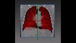
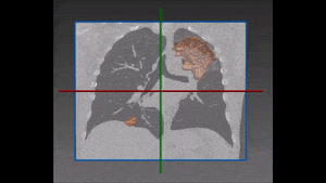
At the first stage they use multi-atlas label fusion to generate priors for anatomical structures and use a simple learning classifier to generate the lymph node candidates. Specifically, they use an SVM classifier to generate candidates in the lungs and a Random forest classifier to generate them in the abdomen.
At the second stage, a 5-layers Convolutional Neural Network (CNN) is used to classify the candidate voxels. There are two main ideas introduced in this part. The first is the combination of the three orthogonal slices (axial, coronal and sagittal) into three channels resembling the RGB channels of color images, which allows the CNN to infer additional information from the relations between them. The second idea is to expand the dataset using rescaling, translations and rotation of the voxels in question.

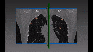
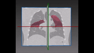
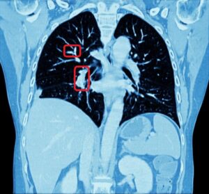
Results of the pulmonary lymph nodes detection

The results of this approach can be described as no less than a drastic improvement over current state-of-the-art results. While the best sensitivity results previously proposed for abdomen and mediastinum lymph node detection were slightly above 70% and 50% respectively, this new approach offers 15% to 20% higher sensitivity, with a much lower ratio of false positives per voxel.
In a subsequent experiment Holger R. Roth et al. also showed that this same architecture, when using both mediastinum and abdomen candidates to teach the CNN, can even reach a result of about 80% sensitivity, with only three false positives per voxel for mediastinum lung lymph nodes detection.
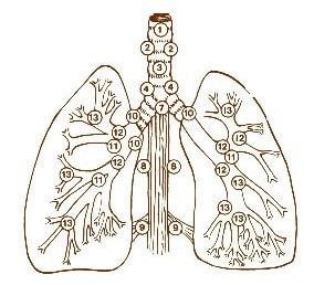
Image courtesy of Manual for Staging of Cancer, 3rd edition, American Joint Committee on Cancer. J B Lippincott Co., 1988.
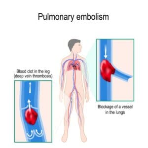
Share

Share on linkedin
Share on twitter
Share on facebook

Main Field

Pulmonology

RSIP Vision’s image processing expertise in medical imaging is currently used in numerous projects, including many applications of computer vision in pulmonology. We are very proud of our contribution to lung medicine: feature detection, lung segmentation and countless other works in the pulmonary imaging field enabled our clients to give the means to physicians to save lives by giving faster and more appropriate treatment to all kind of lung diseases. You can read below about some of our breakthrough developments in Pulmonology and Bronchoscopy.

View Pulmonology

Categories

  • Pulmonology, RSIP Vision Learns

Related Content

Pulmonary embolism

Detecting Pulmonary Embolism from CT Scan

Lung vasculature segmentation

Lung tumors

Lung Tumor Segmentation

Lung Nodules Segmentation

Lung fissures

Lung Fissures Segmentation

Visible lung cancer on CT scan of chest and abdomen

Chest CT Scan Analysis with Deep Learning

Pulmonary embolism

Detecting Pulmonary Embolism from CT Scan

Lung vasculature segmentation

Lung tumors

Lung Tumor Segmentation

Lung Nodules Segmentation

Lung fissures

Lung Fissures Segmentation

Visible lung cancer on CT scan of chest and abdomen

Chest CT Scan Analysis with Deep Learning

Show all

RSIP Vision

Field-tested software solutions and custom R&D, to power your next medical products with innovative AI and image analysis capabilities.

Read more about us

Get in touch

Please fill the following form and our experts will be happy to reply to you soon

Recent News

IBD Scoring – Clario, GI Reviewers and RSIP Vision Team Up

RSIP Neph Announces a Revolutionary Intra-op Solution for Partial Nephrectomy Surgeries

Announcement – RSIP Vision Presents Successful Preliminary Results from Clinical Study of 2D-to-3D Knee Bones Reconstruction

Announcement – New Urological AI Tool for 3D Reconstruction of the Ureter

All news
Upcoming Events
Stay informed for our next events
Subscribe to Our Magazines

Subscribe now and receive the Computer Vision News Magazine every month to your mailbox

 
Subscribe for free
Follow us
Linkedin Twitter Facebook Youtube

contact@rsipvision.com

Terms of Use

Privacy Policy

© All rights reserved to RSIP Vision 2023

Created by Shmulik

  • Our Work
    • title-1
      • Ophthalmology
      • Uncategorized
      • Ophthalmology
      • Pulmonology
      • Cardiology
      • Orthopedics
    • Title-2
      • Orthopedics
  • Success Stories
  • Insights
  • The company