Skip to content
  • Our Work
    • Fields
      • Cardiology
      • ENT
      • Gastro
      • Orthopedics
      • Ophthalmology
      • Pulmonology
      • Surgical
      • Urology
      • Other
    • Modalities
      • Endoscopy
      • Medical Segmentation
      • Microscopy
      • Ultrasound
  • Success Stories
  • Insights
    • Magazine
    • Upcoming Events
    • Webinars
    • Meetups
    • News
    • Blog
  • The company
    • About us
    • Careers
Menu
  • Our Work
    • Fields
      • Cardiology
      • ENT
      • Gastro
      • Orthopedics
      • Ophthalmology
      • Pulmonology
      • Surgical
      • Urology
      • Other
    • Modalities
      • Endoscopy
      • Medical Segmentation
      • Microscopy
      • Ultrasound
  • Success Stories
  • Insights
    • Magazine
    • Upcoming Events
    • Webinars
    • Meetups
    • News
    • Blog
  • The company
    • About us
    • Careers
Contact

Lung vessel segmentation

Blood vessel segmentation of the lungs can help to identify important pulmonary diseases: it contributes to characterize nodules in the lungs, detect pulmonary emboli and evaluate the lungs vasculature in pulmonary hypertension. The patient’s individual values can be compared with normative values to detect and assess the occurrence, the extent and the clinical evolution of the disease. Lung vessel segmentation also helps to achieve the definition of a fissure in the cases where the CT image of the fissure is incomplete.
Lung vessel segmentation

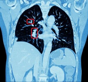
Modern Computed Tomography technology enables entire scans of the lung with submillimeter voxel precision. However, manual segmentation of the complex vessel tree structure is not only an extenuatingly long task for the human; it can also be considered an almost impossible mission for several reasons: first, the boundaries of a vessel (especially the thin ones) are quite difficult to determine in a consistent way due to image noise; second, manual measurements of the vessels in order to assess diameters and branching angles via 2D images are unreliable, mainly because of problems of foreshortening in projected view. Therefore, highly automated segmentation of the pulmonary vascular tree based on 3D image analysis plays a fundamental role in detecting and characterizing the vessel structure.

Automated lung vessels segmentation

As a consequence, the automated segmentation of the vessel tree structure of the lung was understandably considered by our client a crucial process in order to obtain an optimal interpretation of pulmonary CT scans.
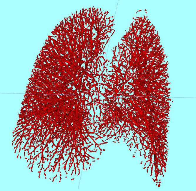
The lung vessel segmentation solution which we provided to our client focused on a complex set of techniques, the core of which is centered on the concept of strain energy vesselness and the use of vessel enhancement filters.
These filters derive structural information from a Hessian matrix computed using Gaussian derivatives. The role of the matrix is to help identify local forms in a specific region of interest. In this case, the targets of the lung vessel enhancement are cross-sections of tubular objects, the round forms of which correspond to the location of blood vessels.
Three invariants are then measured on the values of the Hessian matrix: intensity contrast, structure strength and shape. The purpose of these values is to exclude undesired forms (like lung boundaries) from the image structure and to identify continuous forms bearing consistent intensity to locate the vessel which compose the extracted vascular tree. In addition, the method has also helped the client to identify sparse or void regions of the vascular tree, which correspond to the horizontal and the oblique fissures of the lung.
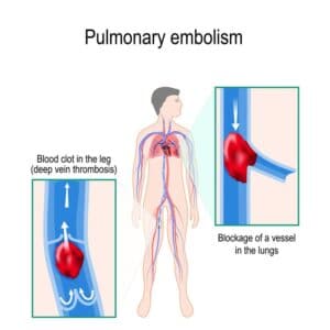
We intentionally decided to test our method against a set of data which presents statistical challenges: respiratory diseases like emphysema, pulmonary nodules or embolisms affect the lungs in a way that introduces additional difficulties to the lung vessel segmentation. The method has proved practical and reliable: the results show that this technique of automatic pulmonary vessel segmentation completes very effectively the vessel tree structure provided by the CT scans of the lung, in such a way that the resulting image is more precise and matchlessly faster than any tentative manual segmentation could be.

Share

Share on linkedin
Share on twitter
Share on facebook

Main Field

Pulmonology

RSIP Vision’s image processing expertise in medical imaging is currently used in numerous projects, including many applications of computer vision in pulmonology. We are very proud of our contribution to lung medicine: feature detection, lung segmentation and countless other works in the pulmonary imaging field enabled our clients to give the means to physicians to save lives by giving faster and more appropriate treatment to all kind of lung diseases. You can read below about some of our breakthrough developments in Pulmonology and Bronchoscopy.

View Pulmonology

Categories

  • Pulmonology, RSIP Vision Learns

Related Content

Pulmonary embolism

Detecting Pulmonary Embolism from CT Scan

Lung vasculature segmentation

Lung tumors

Lung Tumor Segmentation

Lung Nodules Segmentation

Lung fissures

Lung Fissures Segmentation

Visible lung cancer on CT scan of chest and abdomen

Chest CT Scan Analysis with Deep Learning

Pulmonary embolism

Detecting Pulmonary Embolism from CT Scan

Lung vasculature segmentation

Lung tumors

Lung Tumor Segmentation

Lung Nodules Segmentation

Lung fissures

Lung Fissures Segmentation

Visible lung cancer on CT scan of chest and abdomen

Chest CT Scan Analysis with Deep Learning

Show all

RSIP Vision

Field-tested software solutions and custom R&D, to power your next medical products with innovative AI and image analysis capabilities.

Read more about us

Get in touch

Please fill the following form and our experts will be happy to reply to you soon

Recent News

IBD Scoring – Clario, GI Reviewers and RSIP Vision Team Up

RSIP Neph Announces a Revolutionary Intra-op Solution for Partial Nephrectomy Surgeries

Announcement – RSIP Vision Presents Successful Preliminary Results from Clinical Study of 2D-to-3D Knee Bones Reconstruction

Announcement – New Urological AI Tool for 3D Reconstruction of the Ureter

All news
Upcoming Events
Stay informed for our next events
Subscribe to Our Magazines

Subscribe now and receive the Computer Vision News Magazine every month to your mailbox

 
Subscribe for free
Follow us
Linkedin Twitter Facebook Youtube

contact@rsipvision.com

Terms of Use

Privacy Policy

© All rights reserved to RSIP Vision 2023

Created by Shmulik

  • Our Work
    • title-1
      • Ophthalmology
      • Uncategorized
      • Ophthalmology
      • Pulmonology
      • Cardiology
      • Orthopedics
    • Title-2
      • Orthopedics
  • Success Stories
  • Insights
  • The company