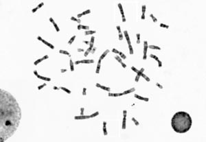
Chromosome classification
Chromosomes are organized structures containing most of living organisms’ DNA. Though important to detect major troubles to an individual’s growth, development and body functioning, the test which identifies and evaluates size, shape and number of chromosomes in the body cells needs human expertise, which is currently very rare. RSIP Vision decided to use convolutional neural networks to perform this chromosomes automated classification with machine learning.
Read More
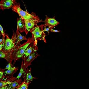
Trajectory tracking of a fluorescent tag
Studying the behavior of bio-molecules and the interaction they have with other molecular structures in their native environment, developing indirect measuring procedures based on tracking of single particle, provides valuable information about processes like viral infections of cells, protein-DNA interactions and other complex biological processes. Analysis of trajectories of a tagged particle is one of many RSIP Vision’s projects tracking objects in a sequence of images with dynamic programming, one of our fields of expertise.
Read More
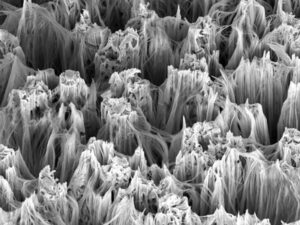
Reconstruction of rough surfaces with shape from focus
Reconstruction of the 3D shape of a surface viewed under the microscope is particularly challenging, owing to the irregular shapes that a surface can take. Irregular surfaces having many sharp bends and peaks have a high frequency texture pattern which needs to be smoothed out through a low-pass filter. The shape-from-focus method provides the framework to do just that, thanks to its ability to stably reconstruct a high frequency surface, as seen in electron microscopy.
Read More
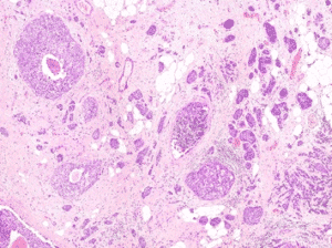
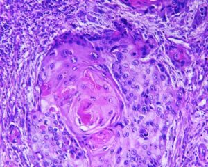
Detecting Mitosis Using Deep Neural Networks
State and progression of breast cancer are assessed through prognostic factors, one of which is the mitotic figure. In a histological sample taken from patients, the fraction of breast tissue cells undergoing replication is used to grade the cancer. RSIP Vision’s algorithms allow fast detection, recognition and classification of the mitotic state of a cell using automatic computational autonomous tools: deep neural networks help distinguish complex patterns in images and finally differentiate between mitotic and non-mitotic cells.
Read More
Automatic segmentation of tumor cells
Molecular analysis of in histology enables quantification of abnormality in a given tissue, assess patient condition, and devise treatment. Tissue samples taken in biopsy allow researchers to screen for therapeutic agents but might not accurately capture the bulk tumor, due to its irregular non-cylindrical shape. This calls for an automated segmentation of tumor cells: RSIP Vision does that in several phases, concluded by machine learning methods which study the cell texture and classify the image accordingly. The end result is a fast and life-saving biopsy scanning and analysis system.
Read More
Bones and Skeleton segmentation
RSIP Vision suggests an automatic segmentation procedure based on iterative binarization of bone tissues density, as observed in Computed Tomography (CT), the most common 3D process used for bone imaging. This method is particularly fast, regardless of whether contrast was used in the CT scans. In fact, images taken with contrast generally display blood with an intensity which is similar to bone; our technique is able to overcome this challenge and to deliver a fast and satisfying bones segmentation and skeleton segmentation solution to our client.
Read More
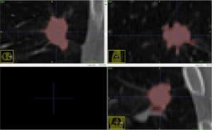
Lungs tumors and nodules segmentation with Deep Learning
It is visually more difficult to identify lung tumors than nodules, since the latter are supposed to have an elliptical shape, while the chromatic aspect of the former is quite hard to distinguish from healthy tissues on a CT image. We use Deep Learning neural networks to overcome this difficulty in a way that is quick to perform, reliable and memory efficient. Our software of computer vision in pulmonology detects and classifies tumors and nodules in the fastest time, to provide our clients a quick and reliable 3D segmentation of lung tumors with Deep Learning.
Read More
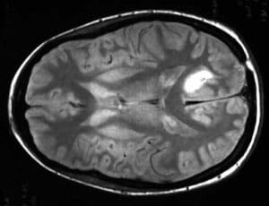
Brain tumor segmentation
In addition to primary tumors, the human brain can also suffer from secondary tumors or brain metastases. The most common cancers that spread from remote areas to the brain are lung, breast, melanoma, kidney, nasal cavity and colon cancers. By the way of segmenting the tumor in the image, brain tumor image processing overcomes anatomical structure challenges. AI-based techniques enable to estimate the volume and spread of the tumor and provide objective and variation-free expected tumor boundaries.
Read More
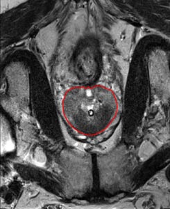
Prostate segmentation in MR images
Prostate cancer is the second most common cancer among American men, with more than 200,000 new cases diagnosed every year and about 1 man in 7 diagnosed during his lifetime. Volume is a key indicator of the health of the prostate, revealing key information about the stage of the cancer, the probable prognosis and viable treatment. The rich experience of RSIP Vision enables us to recommend an approach based on a semi-automatic prostate segmentation to give a precise estimate of the prostate volume.
Read More
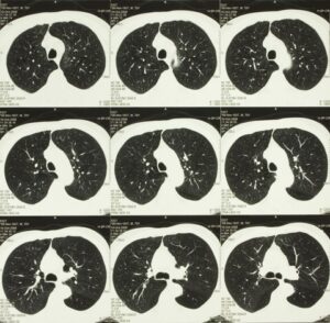
Lung Nodule Classification
Lung cancer early detection is a vital task which is made difficult by the small size of pulmonary nodules, the detection of which on thousands of CT scans every day is excessively time-consuming. Computer-aided lung nodule classification can dramatically boost the speed of diagnosis. Recommended solution starts from bidimensional images obtained from CT scan and displaying suspicious nodules areas: these are inserted into an autoencoder, from which two hundred dimensional features are extracted. These learned features are then confronted with a trained classifier to produce the final lung nodules classification.
Read More
Airways segmentation with Deep Learning
Image processing is a fundamental technique in the quest to identify lung cancer, one of the main causes of death among both men and women, and many other lung pathologies. For the worst diseases, survival rate depends on the stage in which the disease is diagnosed and correct segmentation of airway vessels offers the most effective solution to determine the lesion’s size and location, significantly improving diagnosis and treatment. Our solution is built upon Deep Learning and neural networks.
Read More
Kidney Segmentation
The most dramatically common kidney diseases are: kidney cancer, hitting 50,000 new patients every year only in the U.S.; and kidney failures, which leave the organ unable to remove wastes. Laparoscopic partial nephrectomy operations remove or reduce kidney tumors and some renal malfunctions. We at RSIP Vision help by providing a semi-automatic and very accurate kidney segmentation technique, built on deep learning and neural networks to create a kidney model which would be specific for each patient.
Read More
Pulmonary lobes segmentation
Emphysema quantification and lung nodule detection are among the clinical applications which benefit the most from lobes segmentation in CT scans. Proper lung segmentation is key to determine the boundaries of lobes and prevent pleural damage during examination and treatment. When correctly located, diseases are treated faster and better, hence the call for RSIP Vision to find a faster alternative to time-consuming manual segmentation.
Read More
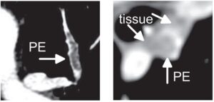
Pulmonary embolism detection
Timely detection of pulmonary embolism via CT angiography is key to reduce mortality risks. Human detection of pulmonary embolism (being often too slow, RSIP Vision recommends a combination of bidimensional and tridimensional image-processing techniques to achieve computer-aided pulmonary embolism detection which enables prompter diagnosis and treatment.
Read More

Date sorting
RSIP Vision has successfully worked in number of dates grading (or dates sorting) projects for our clients. Our automatic fruit recognition system is able to identify with high speed and accuracy all meaningful product features such as size, weight, defect, quality, color, texture, ripeness and others, offering key benefits to our clients: namely, fast and high-volume classification, savings in labor costs, consistent quality and reduced time-to-market.
Read More
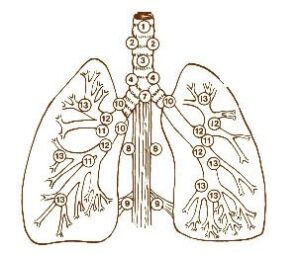
Lung Lymph Nodes Detection
Analyzing pulmonary lymph nodes can give us valuable information for lung cancer diagnosis and treatment. This solution too uses advanced algorithm of computer vision for pulmonology; it also allows to overcome technical difficulties like low image contrast and high nodes variation, offering a drastic improvement over techniques currently used to detect lung lymph nodes.
Read More
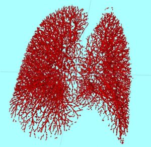
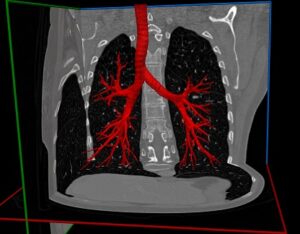
Lung vessel segmentation
Blood vessel segmentation of the lungs can help to identify important pulmonary diseases, characterizing nodules in the lungs, detecting pulmonary emboli and evaluating the lungs vasculature. Our technique of automatic pulmonary vessel segmentation completes very effectively the vessel tree structure provided by the CT scans of the lung, in such a way that the resulting image is more precise and matchlessly faster than any manual segmentation could be.
Read More
Measurement of Retinal Thickness
Retinal thickness is a key measurement used to assess the health of the retina and whether it needs any treatment. Thickness measures can be compared to optimal ranges or to data from the same patient over time, helping ophthalmologists to identify retinal disorders. Advanced optimization methods, borrowed from graph theory, enable us to solve the complex challenge of measuring retinal thickness within reasonable processing time.
Read More
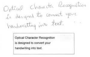
Deep Learning For OCR
OCR used for the visual inspection of documents has found wide application in both industry and research, though it is more commonly found in connection with printed characters than for handwritten ones, mainly owing to the variability in handwritten characters’ shapes and styles. Hence the need for automatic recognition performed by vision-based tailor-made algorithms and adjustment. Deep Neural Networks as a learning mechanism to perform recognition have proved to be particularly powerful tools, due to their high accuracy in both spotting text region and deciphering the characters.
Read More
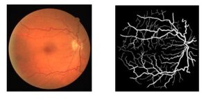
Vessel Segmentation Using Deep Learning
Various segmentation methods, whether based on Convolution Neural Networks or traditional image processing techniques, can be used to delineate the vascular tree in clinical imaging. Given the few features distinguishing veins from arteries (usually brighter and thinner than veins), the challenge consists of training a binary classifier assigning each pixel to the category of vein or artery. This article covers the advantages of using CNNs and deep neural networks for the classification and segmentation of vessels in fundus images.
Read More