Skip to content
  • Our Work
    • Fields
      • Cardiology
      • ENT
      • Gastro
      • Orthopedics
      • Ophthalmology
      • Pulmonology
      • Surgical
      • Urology
      • Other
    • Modalities
      • Endoscopy
      • Medical Segmentation
      • Microscopy
      • Ultrasound
  • Success Stories
  • Insights
    • Magazine
    • Events and Webinars
    • News
    • Blog
  • The company
    • About us
    • Careers
Menu
  • Our Work
    • Fields
      • Cardiology
      • ENT
      • Gastro
      • Orthopedics
      • Ophthalmology
      • Pulmonology
      • Surgical
      • Urology
      • Other
    • Modalities
      • Endoscopy
      • Medical Segmentation
      • Microscopy
      • Ultrasound
  • Success Stories
  • Insights
    • Magazine
    • Events and Webinars
    • News
    • Blog
  • The company
    • About us
    • Careers
Contact

Tag: Lesion detection

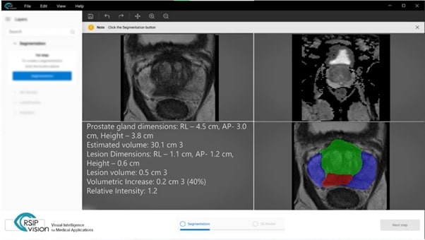
Announcement – New AI Tool for Prostate MRI Analysis to Support PI-RADS Scoring

RSIP Vision Presents New AI Tool for Prostate MRI Analysis to Support PI-RADS Scoring Innovative technology performs automatic segmentation and lesion detection in prostate MRI

Read More
Ultrasound segmentation with AI

Announcement – RSIP Vision introduces an innovative set of AI modules for enhanced medical ultrasound applications

RSIP Vision introduces an innovative set of AI modules for enhanced medical ultrasound applications. These innovative modules empower a wide range of medical applications by

Read More
Airways Segmentation with AI

Announcement – RSIP Vision Launches a Pioneering AI Suite Providing Optimal Solutions to Key Tasks in Lung Surgery

RSIP Vision Launches a Pioneering AI Suite Providing Optimal Solutions to Key Tasks in Lung Surgery. New technology offers critical information enabling pulmonary surgeons to

Read More
Pharma - Tissue Analysis

Tissue Analysis with AI

New AI technologies by RSIP Vision are very powerful in analysis of tissues and histopathology. This complex task, which has been haunting for years the medical community, has now a very practical solution: deep learning gives very fruitful results to several challenges, like the segmentation of cells and nucleus and the classification of the cells according to the detected pathologies.

Read More
RegNet

Deep Learning in Pulmonology

Deep learning has been successfully applied in various applications in pulmonary imaging, including CT registration, airway mapping, real time catheter navigation, and pulmonary nodule detection.

Read More
Giant Retinal Tear

Retinal Detachment Detection

Retinal detachment occurs when part of the retina detaches itself from the pigmented cell layer of the RPE, depriving itself of blood and nutrition: this

Read More
Optical coherence tomography angiography

Optical Coherence Tomography Angiography

Optical coherence tomography angiography uses OCT to produce high-resolution images of the vascular tree in the eye. A fast and non-invasive procedure, it generates high resolution tridimensional images. Stitching multiple images together and increasing image information and clarity, RSIP Vision’s solution allows physicians to quickly and accurately detect pathologies related to vascularization without side effects or clinical hazards.

Read More
Fluorescein Angiogram

Stabilization in fluorescein angiography

One of the most common tests in ophthalmology is fluorescein angiography: fluorescein is injected in the blood and it moves immediately through the blood vessels

Read More
Catheter measurement in angiography

Automatic Catheter Orientation Measurement

Catheters are inserted with measurement equipment at their tips, in order to scan their immediate surroundings. While orientation of the catheter’s tip is unknown throughout insertion, RSIP Vision has employed advanced algorithmic techniques to provide an exact measurement of catheter orientation during angiography, enabling the physician to ascertain the orientation of the catheter’s tip from x-ray images.

Read More
Quantitative Coronary Analysis

Quantitative Coronary Analysis

The main contribution of Quantitative Coronary Analysis (QCA) consists in measuring the diameter of arteries. Angiograms provide coronary images of region suspected of lesions using which our advanced algorithms for vessel detection and segmentation measure the segmented artery’s diameter. Abnormal values (as compared to a constructed reference diameter) are suspected as stenosis. Our system extracts and displays relevant values to the view of medical professionals and their patients.

Read More
Cyst detection

Finding Cysts, Part Five: Final Detection

The goal is to automatically detect the appearance of Cystoid Macular Edema (CME) in Optical Coherence Tomography (OCT) images. The deep learning technique used, Convolutional Neural Networks, takes as an input patches of pixels from within the retina. These patches were generated from previous segmentation of retinal images. A further segmentation of the retina is performed using an image processing algorithm called SLIC. Every superpixel thus generated, after being labeled as in the OCT scan, is fed into the neural network to detect the cyst.

Read More
Layer segmentation of the retina

Finding Cysts Part Three: Layer Segmentation

A series of five articles on our Cysts Detection project using deep learning and Convolutional Neural Networks: 1) our cyst detection method; 2) the cyst denoising process; 3) the retinal layer segmentation; 4) the automatical seed-detection; 5) the final detection of the cysts. Our method is exceptionally successful at finding the cysts themselves and most of their area. Remarkable results are achieved even when using relatively small datasets in the training process.

Read More

Automatic Detection of Macular Cysts

A series of five articles on our Cysts Detection project using deep learning and Convolutional Neural Networks: 1) our cyst detection method; 2) the cyst denoising process; 3) the retinal layer segmentation; 4) the automatical seed-detection; 5) the final detection of the cysts. Our method is exceptionally successful at finding the cysts themselves and most of their area. Remarkable results are achieved even when using relatively small datasets in the training process.

Read More

Get in touch

Please fill the following form and our experts will be happy to reply to you soon

Recent News

Announcement – XPlan.ai Confirms Premier Precision in Peer-Reviewed Clinical Study of its 2D-to-3D Knee Reconstruction Solution

IBD Scoring – Clario, GI Reviewers and RSIP Vision Team Up

RSIP Neph Announces a Revolutionary Intra-op Solution for Partial Nephrectomy Surgeries

Announcement – XPlan.ai by RSIP Vision Presents Successful Preliminary Results from Clinical Study of it’s XPlan 2D-to-3D Knee Bones Reconstruction

All news
Upcoming Events
Stay informed for our next events
Subscribe to Our Magazines

Subscribe now and receive the Computer Vision News Magazine every month to your mailbox

 
Subscribe for free
Follow us
Linkedin Twitter Facebook Youtube

contact@rsipvision.com

Terms of Use

Privacy Policy

© All rights reserved to RSIP Vision 2023

Created by Shmulik

  • Our Work
    • title-1
      • Ophthalmology
      • Uncategorized
      • Ophthalmology
      • Pulmonology
      • Cardiology
      • Orthopedics
    • Title-2
      • Orthopedics
  • Success Stories
  • Insights
  • The company