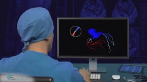
Quantitative Coronary Analysis
Quantitative Coronary Analysis (QCA) refers to the set of methods used to measure the diameter of arteries. The set of tools and algorithms for QCA have been developed for the purpose of lesion and stenosis detection and analysis, and promote rational clinical decision making, risk assessment, and stent placement.

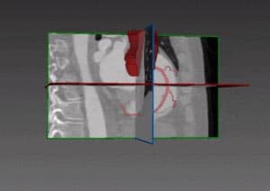
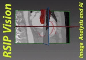
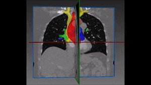
Coronary images in the region suspected of lesions or stenosis are provided by angiograms, which are visualizations of the vascular tree. During the procedure of stenosis detection, images are presented to a doctor, who marks an artery segment based on where the problem lies. Advanced algorithms for vessel detection and segmentation are used to measure the segmented artery’s diameter. An important indication is the point where the artery’s diameter is minimal, which is the suspected position for stenosis.
RSIP Vision has developed an advanced semi-interactive algorithm for quantitative coronary analysis. Using our system a technician or a doctor marks two points along an artery in the image. After which, our adaptation of the Bellman-Ford algorithm is utilized to detect the artery’s center-line and edges. Using a sophisticated segmentation procedure, our system can provide the radius at each point for every given artery.
Points suspected as stenosis are those with abnormal diameter values. Abnormality is determined by constructing a reference diameter (the white line in the attached picture). The reference diameter is simply a linear interpolation between the diameters at the start/end point of the segment. The point where the artery is narrowest compared to a “healthy” artery (the reference) is marked.
More information and measurements are readily available using our system. The minimum and maximum artery diameters and the diameter along the arc of the artery can then be extracted and displayed in an intuitive manner for medical professionals and their patients.
 Cardiology