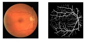
Vessel Segmentation Using Deep Learning
Animal Monitoring With Pattern Recognition
Automatic Identification of Pigs in a Pen Using Pattern Recognition The growing demand for animal products is characterized, at the farmer’s end, by an
On-Combine, Multi-Sensor Environmental Data Collection
Article Summary: On-Combine, Multi-Sensor Data Collection for Post-harvest Assessment of Environmental Stress in Wheat Continuing our series examining interesting articles in the field of computer
Applications in Precision Agriculture
Image Processing Applications in Precision Agriculture In this page, you will learn about image processing applications for precise agriculture. If you want to boost your
Indoor Scene Structure Analysis
Summary: Indoor Scene Structure Analysis for Single Image Depth Estimation This is the first of our series of summaries of interesting texts on computer
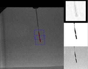
Automatic Catheter Orientation Measurement
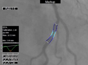
Quantitative Coronary Analysis
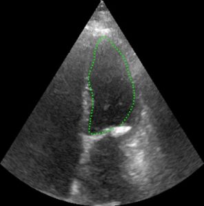
Right Atrium Measurement with Ultrasound
Right Atrium Measurement in Ultrasound Videos Atrial fibrillation is an irregular rhythmic beating of the heart associated with coronary heart disease, high blood pressure
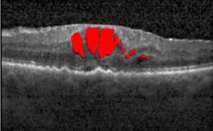
Finding Cysts, Part Five: Final Detection
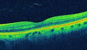
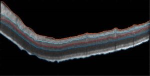
Explaining OCT Scans
What are OCT Scans? Optical coherence tomography (OCT) is a non-invasive imaging method, which produces high-resolution volumetric histological images of tissue. To penetrate deep into biological
Finding Cysts Part Three: Layer Segmentation
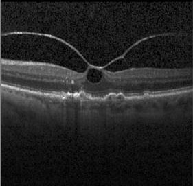
Finding Cysts, Part Two: The Denoising Process
Automatic Detection of Macular Cysts
Defining the Borders within Computer Vision
What’s the Difference between Computer Vision, Image Processing and Machine Learning? In this page, you will learn about Machine Vision, Computer Vision and Image Processing. If you
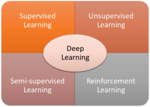
Exploring Deep Learning & CNNs
Deep Learning and Convolutional Neural Networks: RSIP Vision Blogs In this page, you will learn about Computer Vision, Machine Vision and Image Processing. If