Skip to content
  • Our Work
    • Fields
      • Cardiology
      • ENT
      • Gastro
      • Orthopedics
      • Ophthalmology
      • Pulmonology
      • Surgical
      • Urology
      • Other
    • Modalities
      • Endoscopy
      • Medical Segmentation
      • Microscopy
      • Ultrasound
  • Success Stories
  • Insights
    • Magazine
    • Upcoming Events
    • Webinars
    • Meetups
    • News
    • Blog
  • The company
    • About us
    • Careers
Menu
  • Our Work
    • Fields
      • Cardiology
      • ENT
      • Gastro
      • Orthopedics
      • Ophthalmology
      • Pulmonology
      • Surgical
      • Urology
      • Other
    • Modalities
      • Endoscopy
      • Medical Segmentation
      • Microscopy
      • Ultrasound
  • Success Stories
  • Insights
    • Magazine
    • Upcoming Events
    • Webinars
    • Meetups
    • News
    • Blog
  • The company
    • About us
    • Careers
Contact

Chest CT Scan Analysis with Deep Learning

Chest radiography, with modalities such as X-Ray and CT, is now the common practice for the detection and analysis of the progression of lung tumors, tuberculosis and other pulmonary abnormalities. To date, most analysis are done by expert radiographers, who analyze resulting scans and estimate patient prognosis. However, such manual labor is prone to subjectivity and it is time consuming, which hampers large scale fast analysis. This last point poses as a bottleneck for retrospective studies on a database of chest scans aiming at finding disease characteristics.
Visible lung cancer on CT scan of chest and abdomen

Automated Chest CT Scan Analysis

Computer-aided automated analysis of chest scans can greatly alleviate the downsides of manual analysis, by providing fast, accurate and reproducible detection of lung abnormalities.

With the advances in neural networks and deep learning methodologies, it is now relatively easy to detect dormant lung abnormalities and follow them over successive acquisitions up to disease manifestation. However, prior to the use of an automated image classifier, such as convolutional neural networks, image preprocessing must take place to ensure features extraction is performed in designated position on the lungs.

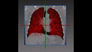
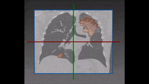
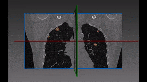
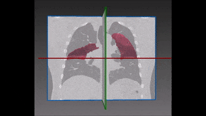
Analysis towards automated pulmonary disease detection is based on two major steps. At the first stage, the left and right lung are to be separated in the image from the surrounding thoracic tissue. This stage by itself can pose several technical challenges due to variation in lung shapes, patient orientation and cropped X-ray images, while a failure to correctly delineate lung boundaries can result in false information being processed. Several practices exist for the extraction of lung shapes: some of the most common use lung image masks, active contours, and machine-learning based methods. Care must be taken in the boundary detection as abnormalities oftentimes appear in graytone levels similar to those of lung boundaries, with the result of possibly skewing the latter.

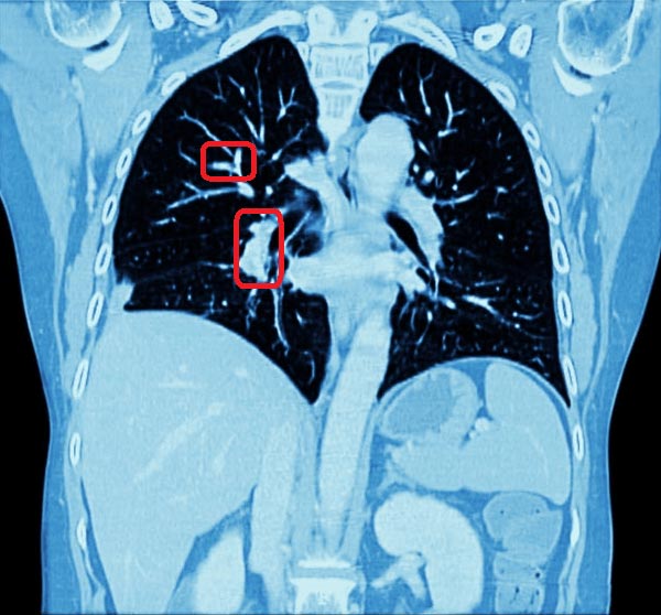
Once lung boundaries are extracted, a second stage examines the characteristics of the lung region within the image. Classification of abnormalities based on morphologies, position and gray levels, is routinely done by radiographers. This knowledge can be translated into an autonomous machine decision procedure by training a convolutional neural network to discern between pathologies (a classifier), based on local image characteristics.

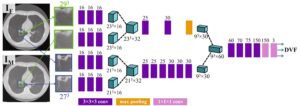
Convolutional neural network consists of image convolutional layers to process an image (or regions of), where information between different layers is passed and processed according to a predefined architecture, most often identified empirically. The identification of network architecture for optimal classification is one of the challenges faced by machine learning experts and is subject to the experience of the engineers and the complexity of the problem at hand.

RSIP Vision is a global leading expert in tailor-made machine learning and computer vision algorithms. Our clients around the globe enjoy our accumulated knowledge and experience, gathered from several decades of experience. To get consulting for your project contact us today. To learn more about RSIP Vision’s algorithmic solutions, visit our project page now.

Share

Share on linkedin
Share on twitter
Share on facebook

Main Field

Pulmonology

RSIP Vision’s image processing expertise in medical imaging is currently used in numerous projects, including many applications of computer vision in pulmonology. We are very proud of our contribution to lung medicine: feature detection, lung segmentation and countless other works in the pulmonary imaging field enabled our clients to give the means to physicians to save lives by giving faster and more appropriate treatment to all kind of lung diseases. You can read below about some of our breakthrough developments in Pulmonology and Bronchoscopy.

View Pulmonology

Categories

  • Pulmonology, RSIP Vision Learns

Related Content

Pulmonary embolism

Detecting Pulmonary Embolism from CT Scan

Lung vasculature segmentation

Lung tumors

Lung Tumor Segmentation

Lung Nodules Segmentation

Lung fissures

Lung Fissures Segmentation

RegNet

Deep Learning in Pulmonology

Pulmonary embolism

Detecting Pulmonary Embolism from CT Scan

Lung vasculature segmentation

Lung tumors

Lung Tumor Segmentation

Lung Nodules Segmentation

Lung fissures

Lung Fissures Segmentation

RegNet

Deep Learning in Pulmonology

Show all

RSIP Vision

Field-tested software solutions and custom R&D, to power your next medical products with innovative AI and image analysis capabilities.

Read more about us

Get in touch

Please fill the following form and our experts will be happy to reply to you soon

Recent News

IBD Scoring – Clario, GI Reviewers and RSIP Vision Team Up

RSIP Neph Announces a Revolutionary Intra-op Solution for Partial Nephrectomy Surgeries

Announcement – RSIP Vision Presents Successful Preliminary Results from Clinical Study of 2D-to-3D Knee Bones Reconstruction

Announcement – New Urological AI Tool for 3D Reconstruction of the Ureter

All news
Upcoming Events
Stay informed for our next events
Subscribe to Our Magazines

Subscribe now and receive the Computer Vision News Magazine every month to your mailbox

 
Subscribe for free
Follow us
Linkedin Twitter Facebook Youtube

contact@rsipvision.com

Terms of Use

Privacy Policy

© All rights reserved to RSIP Vision 2023

Created by Shmulik

  • Our Work
    • title-1
      • Ophthalmology
      • Uncategorized
      • Ophthalmology
      • Pulmonology
      • Cardiology
      • Orthopedics
    • Title-2
      • Orthopedics
  • Success Stories
  • Insights
  • The company