Skip to content
  • Our Work
    • Fields
      • Cardiology
      • ENT
      • Gastro
      • Orthopedics
      • Ophthalmology
      • Pulmonology
      • Surgical
      • Urology
      • Other
    • Modalities
      • Endoscopy
      • Medical Segmentation
      • Microscopy
      • Ultrasound
  • Success Stories
  • Insights
    • Magazine
    • Upcoming Events
    • Webinars
    • Meetups
    • News
    • Blog
  • The company
    • About us
    • Careers
Menu
  • Our Work
    • Fields
      • Cardiology
      • ENT
      • Gastro
      • Orthopedics
      • Ophthalmology
      • Pulmonology
      • Surgical
      • Urology
      • Other
    • Modalities
      • Endoscopy
      • Medical Segmentation
      • Microscopy
      • Ultrasound
  • Success Stories
  • Insights
    • Magazine
    • Upcoming Events
    • Webinars
    • Meetups
    • News
    • Blog
  • The company
    • About us
    • Careers
Contact

Detecting Pulmonary Embolism from CT Scan

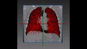
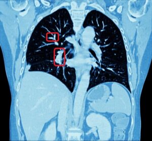
Pulmonary embolism is a very dangerous condition, which happens when a clot of blood moves from somewhere (generally the legs) to the heart and then finds its way to the lungs. As blood vessels become finer and finer, at some point the clot gets stuck there and blocks the supply of blood, with all the dire consequences resulting from that.

Pulmonary embolism

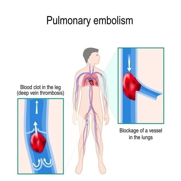
Detecting the clot location is a challenging task, since the description of the pain by the patient is usually not enough precise to enable a clear-cut diagnosis. Often, the emergency room physician will send the patient to a CT scan of the lung, transferring the challenge to the radiologist. A trained radiologist will be able to detect the pulmonary embolism, but this manual process will generally take some time and it might also involve some mistakes.

Detecting Pulmonary Embolism with AI

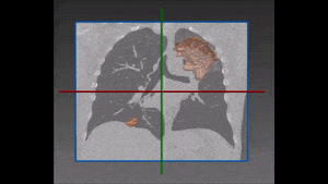
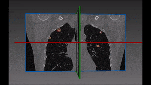
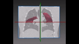
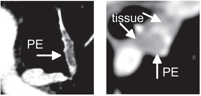
AI technology can give a good solution to this problem, by providing an automated detection of this condition based on several indicators: first, a specific algorithm distinguishes veins from arteries and detects the interested blood vessel, which will always be a vein. In a further step, the algorithm needs to detect the location of the missing vein part that isn’t seen in the CT: in order words, it locates the vein’s part which is invisible in the scan, because the blood is blocked and cannot reach that area. In that case, we use our knowledge of the blood vessels tree structure to evaluate the different densities in the different parts of the lung and only then we will infer that there is one area where blood’s flow is nil or significantly limited. RSIP Vision’s rich experience teaches a combination of several steps is what leads to the right solution: first of all, detection of the blood vessels, then detection of the missing part and segmentation of the lungs; we recommend that some of these tasks parts be performed with Deep Learning algorithms, while other tasks can be done using algorithms taken from graph theory and optimization techniques.

Pulmonary embolism

On the same subject

Share

Share on linkedin
Share on twitter
Share on facebook

Main Field

Pulmonology

RSIP Vision’s image processing expertise in medical imaging is currently used in numerous projects, including many applications of computer vision in pulmonology. We are very proud of our contribution to lung medicine: feature detection, lung segmentation and countless other works in the pulmonary imaging field enabled our clients to give the means to physicians to save lives by giving faster and more appropriate treatment to all kind of lung diseases. You can read below about some of our breakthrough developments in Pulmonology and Bronchoscopy.

View Pulmonology

Categories

  • Pulmonology, RSIP Vision Learns

Related Content

Lung vasculature segmentation

Lung tumors

Lung Tumor Segmentation

Lung Nodules Segmentation

Lung fissures

Lung Fissures Segmentation

Visible lung cancer on CT scan of chest and abdomen

Chest CT Scan Analysis with Deep Learning

RegNet

Deep Learning in Pulmonology

Lung vasculature segmentation

Lung tumors

Lung Tumor Segmentation

Lung Nodules Segmentation

Lung fissures

Lung Fissures Segmentation

Visible lung cancer on CT scan of chest and abdomen

Chest CT Scan Analysis with Deep Learning

RegNet

Deep Learning in Pulmonology

Show all

RSIP Vision

Field-tested software solutions and custom R&D, to power your next medical products with innovative AI and image analysis capabilities.

Read more about us

Get in touch

Please fill the following form and our experts will be happy to reply to you soon

Recent News

IBD Scoring – Clario, GI Reviewers and RSIP Vision Team Up

RSIP Neph Announces a Revolutionary Intra-op Solution for Partial Nephrectomy Surgeries

Announcement – RSIP Vision Presents Successful Preliminary Results from Clinical Study of 2D-to-3D Knee Bones Reconstruction

Announcement – New Urological AI Tool for 3D Reconstruction of the Ureter

All news
Upcoming Events
Stay informed for our next events
Subscribe to Our Magazines

Subscribe now and receive the Computer Vision News Magazine every month to your mailbox

 
Subscribe for free
Follow us
Linkedin Twitter Facebook Youtube

contact@rsipvision.com

Terms of Use

Privacy Policy

© All rights reserved to RSIP Vision 2023

Created by Shmulik

  • Our Work
    • title-1
      • Ophthalmology
      • Uncategorized
      • Ophthalmology
      • Pulmonology
      • Cardiology
      • Orthopedics
    • Title-2
      • Orthopedics
  • Success Stories
  • Insights
  • The company