Skip to content
  • Our Work
    • Fields
      • Cardiology
      • ENT
      • Gastro
      • Orthopedics
      • Ophthalmology
      • Pulmonology
      • Surgical
      • Urology
      • Other
    • Modalities
      • Endoscopy
      • Medical Segmentation
      • Microscopy
      • Ultrasound
  • Success Stories
  • Insights
    • Magazine
    • Upcoming Events
    • Webinars
    • Meetups
    • News
    • Blog
  • The company
    • About us
    • Careers
Menu
  • Our Work
    • Fields
      • Cardiology
      • ENT
      • Gastro
      • Orthopedics
      • Ophthalmology
      • Pulmonology
      • Surgical
      • Urology
      • Other
    • Modalities
      • Endoscopy
      • Medical Segmentation
      • Microscopy
      • Ultrasound
  • Success Stories
  • Insights
    • Magazine
    • Upcoming Events
    • Webinars
    • Meetups
    • News
    • Blog
  • The company
    • About us
    • Careers
Contact

Lung Nodules Segmentation

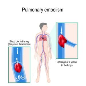
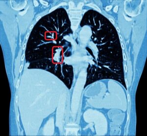
Pulmonary nodules (AKA lung nodules) are small masses (up to 30mm) of tissue surrounded by pulmonary parenchyma. They are quite common finding on computerized tomography (CT) scans, and although most lung nodules are benign, some are cancerous. Some of the characteristics of the nodules may indicate high suspicion of malignancy such as large size, irregular/spiculated borders or inhomogeneous density. In that case, a tissue biopsy (percutaneous or via bronchoscopy) might be indicated.

Lung nodules segmentation

Nodules volume and borders assessment are important in cancer diagnosis and staging. CT offers very reliable imaging for evaluation and follow-up of nodule size, growth, and location. It also allows visualization of nodule attenuation (density) and borders.

Nodule segmentation can also assist the physician during the biopsy, whether it is done via bronchoscopy or percutaneously, by helping the physician choose the best trajectory, hence minimizing complications.

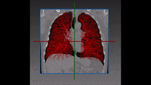
Automated lung nodules segmentation

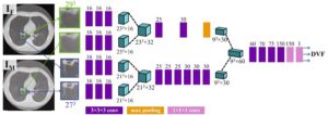
RSIP Vision has developed a state-of-the-art module for detection of lung nodules. Traditional methods based on classic grey level based techniques would not work in this challenging task. The AI methods were used to address various critical issues to distinguish between nodules and blood vessels that have very similar appearance in grey scale. Hence, the appearance of nodules might be very different between the solid ones and the Ground-Glass opacity ones.

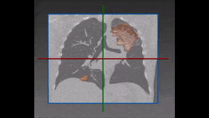
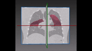
The Deep Learning methods are very versatile and include powerful methods like U-Net and Mask-R CNN that can derive maximum information from the scan in the 3D and the environment of the nodule as well. Much care is taken to furnish the most precise annotated images to the system. During the training phase, the weights of the network get adjusted and refined for the specific task at hand. The result is a very accurate lung nodules segmentation with Deep Learning, that can give you much better results than the ones you currently have. Contact now our engineers to learn more about the automated pulmonary nodules segmentation!

Share

Share on linkedin
Share on twitter
Share on facebook

Main Field

Pulmonology

RSIP Vision’s image processing expertise in medical imaging is currently used in numerous projects, including many applications of computer vision in pulmonology. We are very proud of our contribution to lung medicine: feature detection, lung segmentation and countless other works in the pulmonary imaging field enabled our clients to give the means to physicians to save lives by giving faster and more appropriate treatment to all kind of lung diseases. You can read below about some of our breakthrough developments in Pulmonology and Bronchoscopy.

View Pulmonology

Categories

  • Pulmonology, RSIP Vision Learns

Related Content

Pulmonary embolism

Detecting Pulmonary Embolism from CT Scan

Lung vasculature segmentation

Lung tumors

Lung Tumor Segmentation

Lung fissures

Lung Fissures Segmentation

Visible lung cancer on CT scan of chest and abdomen

Chest CT Scan Analysis with Deep Learning

RegNet

Deep Learning in Pulmonology

Show all

RSIP Vision

Field-tested software solutions and custom R&D, to power your next medical products with innovative AI and image analysis capabilities.

Read more about us

Get in touch

Please fill the following form and our experts will be happy to reply to you soon

Recent News

IBD Scoring – Clario, GI Reviewers and RSIP Vision Team Up

RSIP Neph Announces a Revolutionary Intra-op Solution for Partial Nephrectomy Surgeries

Announcement – RSIP Vision Presents Successful Preliminary Results from Clinical Study of 2D-to-3D Knee Bones Reconstruction

Announcement – New Urological AI Tool for 3D Reconstruction of the Ureter

All news
Upcoming Events
Stay informed for our next events
Subscribe to Our Magazines

Subscribe now and receive the Computer Vision News Magazine every month to your mailbox

 
Subscribe for free
Follow us
Linkedin Twitter Facebook Youtube

contact@rsipvision.com

Terms of Use

Privacy Policy

© All rights reserved to RSIP Vision 2023

Created by Shmulik

  • Our Work
    • title-1
      • Ophthalmology
      • Uncategorized
      • Ophthalmology
      • Pulmonology
      • Cardiology
      • Orthopedics
    • Title-2
      • Orthopedics
  • Success Stories
  • Insights
  • The company