Skip to content
  • Our Work
    • Fields
      • Cardiology
      • ENT
      • Gastro
      • Orthopedics
      • Ophthalmology
      • Pulmonology
      • Surgical
      • Urology
      • Other
    • Modalities
      • Endoscopy
      • Medical Segmentation
      • Microscopy
      • Ultrasound
  • Success Stories
  • Insights
    • Magazine
    • Upcoming Events
    • Webinars
    • Meetups
    • News
    • Blog
  • The company
    • About us
    • Careers
Menu
  • Our Work
    • Fields
      • Cardiology
      • ENT
      • Gastro
      • Orthopedics
      • Ophthalmology
      • Pulmonology
      • Surgical
      • Urology
      • Other
    • Modalities
      • Endoscopy
      • Medical Segmentation
      • Microscopy
      • Ultrasound
  • Success Stories
  • Insights
    • Magazine
    • Upcoming Events
    • Webinars
    • Meetups
    • News
    • Blog
  • The company
    • About us
    • Careers
Contact

Lung Tumor Segmentation

Lung cancer is the most common cancer related mortality cause among men, and second in women worldwide. Primary lung cancer is usually divided into two groups: Non-Small Cell Lung Cancer (NSCLC), and Small Cell Lung Cancer (SCLC), the former being the most common. In cases that are diagnosed late, prognosis might be grim with high mortality rates. However, early diagnosis and treatment can improve outcome. The lung is also a common site for metastatic disease. Common primary tumors which spread to the lungs, include colorectal cancer, breast cancer, renal cancer, melanoma and head and neck cancers.Lung tumors

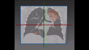
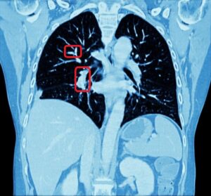
Lung tumors might appear in imaging as nodules (<3cm), or as masses (>3 cm). In order to identify the type of lung tumor, a biopsy is usually performed. Tissue sample can be obtained transbronchial via bronchoscopy, percutaneous guided by CT, thoracoscopically or via open biopsy. Usually after pathological confirmation of malignancy, the disease extent is classified according to the TNM classification of malignant tumors, and helps determine treatment protocol. A measure called Response Evaluation Criteria In Solid Tumors (RECIST) is used to evaluate treatment response.

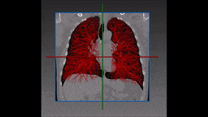
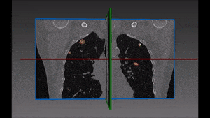
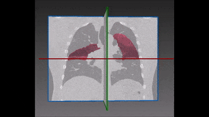
Automated lung tumor segmentation

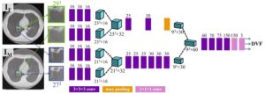
Lung tumor segmentation can assist in diagnosis, staging and follow-up, and can also be used by the physician during biopsy trajectory planning, surgery and image-guided ablation. Segmentation of tumors can be quite challenging, since their shape and size varies a lot. This means, that it is important to gather a large quantity of data to train the deep learning system and also that these data must be accurately annotated by a radiologist. In addition, distinguishing between tumor tissue and other lung findings such as pleural effusion, lymph nodes and consolidation can be difficult due to similar density. In order to deal with this challenge, RSIP Vision has built a proprietary neural network architecture that takes into account both global and local context of the tumor. This cutting edge AI module is today’s best solution for lung tumors segmentation. Contact RSIP Vision’s engineers to learn how.

Share

Share on linkedin
Share on twitter
Share on facebook

Main Field

Pulmonology

RSIP Vision’s image processing expertise in medical imaging is currently used in numerous projects, including many applications of computer vision in pulmonology. We are very proud of our contribution to lung medicine: feature detection, lung segmentation and countless other works in the pulmonary imaging field enabled our clients to give the means to physicians to save lives by giving faster and more appropriate treatment to all kind of lung diseases. You can read below about some of our breakthrough developments in Pulmonology and Bronchoscopy.

View Pulmonology

Categories

  • Pulmonology, RSIP Vision Learns

Related Content

Pulmonary embolism

Detecting Pulmonary Embolism from CT Scan

Lung vasculature segmentation

Lung Nodules Segmentation

Lung fissures

Lung Fissures Segmentation

Visible lung cancer on CT scan of chest and abdomen

Chest CT Scan Analysis with Deep Learning

RegNet

Deep Learning in Pulmonology

Pulmonary embolism

Detecting Pulmonary Embolism from CT Scan

Lung vasculature segmentation

Lung Nodules Segmentation

Lung fissures

Lung Fissures Segmentation

Visible lung cancer on CT scan of chest and abdomen

Chest CT Scan Analysis with Deep Learning

RegNet

Deep Learning in Pulmonology

Show all

RSIP Vision

Field-tested software solutions and custom R&D, to power your next medical products with innovative AI and image analysis capabilities.

Read more about us

Get in touch

Please fill the following form and our experts will be happy to reply to you soon

Recent News

IBD Scoring – Clario, GI Reviewers and RSIP Vision Team Up

RSIP Neph Announces a Revolutionary Intra-op Solution for Partial Nephrectomy Surgeries

Announcement – RSIP Vision Presents Successful Preliminary Results from Clinical Study of 2D-to-3D Knee Bones Reconstruction

Announcement – New Urological AI Tool for 3D Reconstruction of the Ureter

All news
Upcoming Events
Stay informed for our next events
Subscribe to Our Magazines

Subscribe now and receive the Computer Vision News Magazine every month to your mailbox

 
Subscribe for free
Follow us
Linkedin Twitter Facebook Youtube

contact@rsipvision.com

Terms of Use

Privacy Policy

© All rights reserved to RSIP Vision 2023

Created by Shmulik

  • Our Work
    • title-1
      • Ophthalmology
      • Uncategorized
      • Ophthalmology
      • Pulmonology
      • Cardiology
      • Orthopedics
    • Title-2
      • Orthopedics
  • Success Stories
  • Insights
  • The company