Skip to content
  • Our Work
    • Fields
      • Cardiology
      • ENT
      • Gastro
      • Orthopedics
      • Ophthalmology
      • Pulmonology
      • Surgical
      • Urology
      • Other
    • Modalities
      • Endoscopy
      • Medical Segmentation
      • Microscopy
      • Ultrasound
  • Success Stories
  • Insights
    • Magazine
    • Upcoming Events
    • Webinars
    • Meetups
    • News
    • Blog
  • The company
    • About us
    • Careers
Menu
  • Our Work
    • Fields
      • Cardiology
      • ENT
      • Gastro
      • Orthopedics
      • Ophthalmology
      • Pulmonology
      • Surgical
      • Urology
      • Other
    • Modalities
      • Endoscopy
      • Medical Segmentation
      • Microscopy
      • Ultrasound
  • Success Stories
  • Insights
    • Magazine
    • Upcoming Events
    • Webinars
    • Meetups
    • News
    • Blog
  • The company
    • About us
    • Careers
Contact

Lung vasculature segmentation

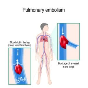
Lungs vasculature has a major part in blood oxygenation. The complicated branches of arteries and veins, accompanied by the intricate bronchial tree are in charge of gas exchange, where inhaled air, containing oxygen, enters the lungs and through the bronchial tree it reaches the alveoli. There, oxygen enters the thin capillaries covering the alveolar sac. In a similar manner, carbon dioxide exits the capillaries into the alveoli and exhaled. From the lung, oxygenated blood reaches the left atrium of the heart via the pulmonary veins. Then, the blood enters the left ventricle and into the aorta, which distributes the oxygenated blood to the rest of the body. Deoxygenated blood is carried from the body to the heart by the vena cava, and travels into the lungs through the pulmonary arteries, where it is later oxygenated as mentioned above.

Lung vessels segmentation

Automated lung vasculature segmentation

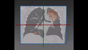
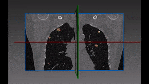
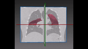
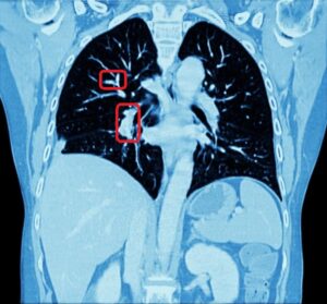
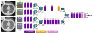
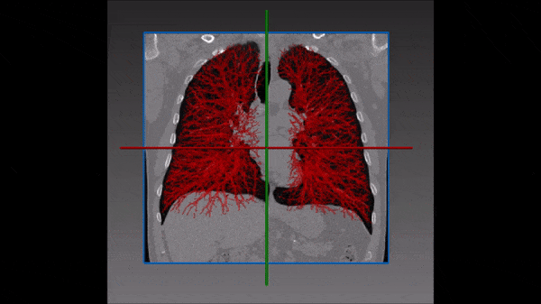
Mapping pulmonary blood vessels, major and small, is essential in clinical setting. For example, knowing the vasculature helps with planning of surgeries within the chest cavity and the lungs, including segmentectomy, lobectomy, pneumonectomy or lung biopsy. In addition, knowledge of the vessels branching may help with distinguishing vessels from nodules, masses or other pathologies involving focal opacities. Furthermore, signs of pulmonary hypertension or  pulmonary embolism can be identified by proper segmentation of the vessels. In the case of an embolism, determining which vessel is occluded could help choosing the best treatment possible. Manual tracing of the pulmonary vessels can be extremely time consuming for the radiologist. Even the experienced physician may come across difficulties when trying to trace the smallest vessels, due to image noise. Another challenge is given by the difficulty in distinguishing between lung vessels and  the tumors which are crossed by them: they have different shape but similar intensity and thus are difficult to segment with classical computer vision techniques. RSIP Vision has developed the most accurate AI module for lung vessel segmentation to solve ideally these challenges. Our automated module uses deep learning neural networks to apply the full power of AI algorithms to lung vasculature segmentation. Contact us and we shall discuss with you the fittest solution for your project.

Share

Share on linkedin
Share on twitter
Share on facebook

Main Field

Pulmonology

RSIP Vision’s image processing expertise in medical imaging is currently used in numerous projects, including many applications of computer vision in pulmonology. We are very proud of our contribution to lung medicine: feature detection, lung segmentation and countless other works in the pulmonary imaging field enabled our clients to give the means to physicians to save lives by giving faster and more appropriate treatment to all kind of lung diseases. You can read below about some of our breakthrough developments in Pulmonology and Bronchoscopy.

View Pulmonology

Categories

  • Pulmonology, RSIP Vision Learns

Related Content

Pulmonary embolism

Detecting Pulmonary Embolism from CT Scan

Lung tumors

Lung Tumor Segmentation

Lung Nodules Segmentation

Lung fissures

Lung Fissures Segmentation

Visible lung cancer on CT scan of chest and abdomen

Chest CT Scan Analysis with Deep Learning

RegNet

Deep Learning in Pulmonology

Show all

RSIP Vision

Field-tested software solutions and custom R&D, to power your next medical products with innovative AI and image analysis capabilities.

Read more about us

Get in touch

Please fill the following form and our experts will be happy to reply to you soon

Recent News

IBD Scoring – Clario, GI Reviewers and RSIP Vision Team Up

RSIP Neph Announces a Revolutionary Intra-op Solution for Partial Nephrectomy Surgeries

Announcement – RSIP Vision Presents Successful Preliminary Results from Clinical Study of 2D-to-3D Knee Bones Reconstruction

Announcement – New Urological AI Tool for 3D Reconstruction of the Ureter

All news
Upcoming Events
Stay informed for our next events
Subscribe to Our Magazines

Subscribe now and receive the Computer Vision News Magazine every month to your mailbox

 
Subscribe for free
Follow us
Linkedin Twitter Facebook Youtube

contact@rsipvision.com

Terms of Use

Privacy Policy

© All rights reserved to RSIP Vision 2023

Created by Shmulik

  • Our Work
    • title-1
      • Ophthalmology
      • Uncategorized
      • Ophthalmology
      • Pulmonology
      • Cardiology
      • Orthopedics
    • Title-2
      • Orthopedics
  • Success Stories
  • Insights
  • The company