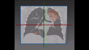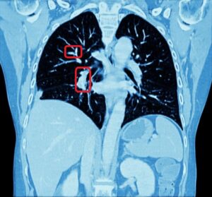

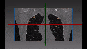
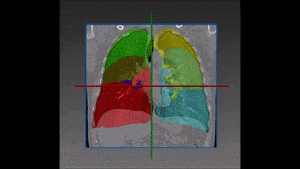
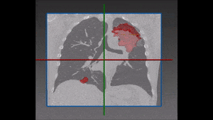
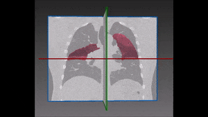
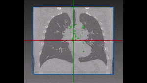
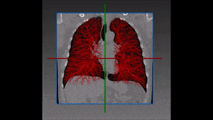
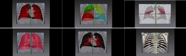
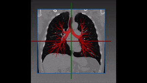
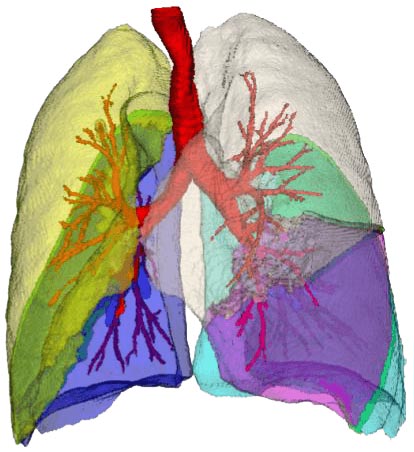

Detecting Pulmonary Embolism from CT Scan
Pulmonary embolism is a very dangerous condition, which happens when a clot of blood moves from somewhere (generally the legs) to the heart and then finds its way to the lungs. As blood vessels become finer and finer, at some point the clot gets stuck there and blocks the supply of blood, with all the dire consequences resulting from that. Detecting the clot location is a challenging task, since the description of the pain by the patient is usually not enough precise to enable a clear-cut diagnosis. Often, the emergency