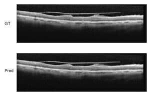
Skip to content
  • Our Work
    • Fields
      • Cardiology
      • ENT
      • Gastro
      • Orthopedics
      • Ophthalmology
      • Pulmonology
      • Surgical
      • Urology
      • Other
    • Modalities
      • Endoscopy
      • Medical Segmentation
      • Microscopy
      • Ultrasound
  • Success Stories
  • Insights
    • Magazine
    • Events and Webinars
    • News
    • Blog
  • The company
    • About us
    • Careers
Menu
  • Our Work
    • Fields
      • Cardiology
      • ENT
      • Gastro
      • Orthopedics
      • Ophthalmology
      • Pulmonology
      • Surgical
      • Urology
      • Other
    • Modalities
      • Endoscopy
      • Medical Segmentation
      • Microscopy
      • Ultrasound
  • Success Stories
  • Insights
    • Magazine
    • Events and Webinars
    • News
    • Blog
  • The company
    • About us
    • Careers
Contact

Explaining OCT Scans

What are OCT Scans?

Optical coherence tomography (OCT) is a non-invasive imaging method, which produces high-resolution volumetric histological images of tissue. To penetrate deep into biological tissues, OCT employs near-infrared light with long wavelength. Ever since its emergence in the early 1990s, both modalities and algorithms for OCT imaging have undergone drastic improvement in acquisition speed and resolution. These advancements have made OCT an established field and the golden standard for histological imaging in ophthalmology. It is through the use of OCT that physicians today routinely diagnose macular diseases such as neovascularization, diabetes, and age-related macular degeneration. OCT is also being used to examine compositions and layers of material in fields such as art and history.

OCT scan

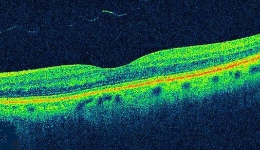
The mechanisms of OCT are similar to that of ultrasound, only instead of sound waves OCT uses the reflection of light. The resolution obtained by OCT is 1-2 orders of magnitude higher than ultrasound, and has been demonstrated to work on both transparent and non-transparent tissue with high reflection. In OCT many one-dimensional scans (a-scans) are performed at several depths to create a two-dimensional image (b-scan). Those b-scans, if acquired closely and rapidly, can be translated into a volumetric image (c-scan) of a retina, for example.

There are three main advantages of OCT over older methods: scan speed, that it is non-invasive, and three-dimensional data generation. With OCT each volumetric scan can take up to 6 seconds in comparison to 10-30 minutes for methods like fluorescein tomography, which produces only two-dimensional images.

However, despite of the speed of OCT, patient motion and blinking might introduce artifacts into imaging, and one must correct for them in order to enjoy the benefits of OCT. Poor fixation of patients’ eyes produces artifacts in measurements, which is common due to the high frequency of macular disease occurrence in elderly patients.  Typically, patient blinking during OCT scanning produces black lines across the image, and deterioration of signal is the result of patient motion. One then has to compensate for patient motion during acquisition by stabilizing the ophthalmic system using eye tracking procedures. RSIP Vision has developed software for eye tracking and stabilization.

The introduction of spectral domain OCT has significantly shortened acquisition time, and has allowed near video rate imaging. This has benefits for real-time eye tracking and calibration of the OCT machine, and several OCT machine vendors have incorporated calibration algorithms in their machines based on tracking. Some examples for common machines are Spectralis, RTVue, and tracking OCT, all systems we at RSIP Vision have worked with.

Share

Share on linkedin
Share on twitter
Share on facebook

Main Field

Ophthalmology

RSIP Vision has developed countless projects in the field of ophthalmology for its clients. Computer vision with sophisticated ophthalmic imaging, measurement techniques and AI can yield better results: great precision, accurate diagnosis and best interventional treatment for many pathologies.

View Ophthalmology

Categories

  • Ophthalmology, RSIP Vision Learns

Related Content

Super-Resolution in OCT images

Super-Resolution in OCT images

Zoom-in-Net

Deep Learning in Ophthalmology

Classification and Segmentation of Dendritic cells

Classification and Segmentation of Dendritic Cells

Alzheimer's Disease - AD

Degenerative Diseases Detection in the Eye

Temporary pediatric strabismus in newborn baby

Image processing for pediatric strabismus

ROP - Vessel tortuosity in Retinopathy of Prematurity

ROP: Retinopathy of Prematurity

Super-Resolution in OCT images

Super-Resolution in OCT images

Zoom-in-Net

Deep Learning in Ophthalmology

Classification and Segmentation of Dendritic cells

Classification and Segmentation of Dendritic Cells

Alzheimer's Disease - AD

Degenerative Diseases Detection in the Eye

Temporary pediatric strabismus in newborn baby

Image processing for pediatric strabismus

ROP - Vessel tortuosity in Retinopathy of Prematurity

ROP: Retinopathy of Prematurity

Show all

RSIP Vision

Field-tested software solutions and custom R&D, to power your next medical products with innovative AI and image analysis capabilities.

Read more about us

Get in touch

Please fill the following form and our experts will be happy to reply to you soon

Recent News

Announcement – XPlan.ai Confirms Premier Precision in Peer-Reviewed Clinical Study of its 2D-to-3D Knee Reconstruction Solution

IBD Scoring – Clario, GI Reviewers and RSIP Vision Team Up

RSIP Neph Announces a Revolutionary Intra-op Solution for Partial Nephrectomy Surgeries

Announcement – XPlan.ai by RSIP Vision Presents Successful Preliminary Results from Clinical Study of it’s XPlan 2D-to-3D Knee Bones Reconstruction

All news
Upcoming Events
Stay informed for our next events
Subscribe to Our Magazines

Subscribe now and receive the Computer Vision News Magazine every month to your mailbox

 
Subscribe for free
Follow us
Linkedin Twitter Facebook Youtube

contact@rsipvision.com

Terms of Use

Privacy Policy

© All rights reserved to RSIP Vision 2023

Created by Shmulik

  • Our Work
    • title-1
      • Ophthalmology
      • Uncategorized
      • Ophthalmology
      • Pulmonology
      • Cardiology
      • Orthopedics
    • Title-2
      • Orthopedics
  • Success Stories
  • Insights
  • The company