Skip to content
  • Our Work
    • Fields
      • Cardiology
      • ENT
      • Gastro
      • Orthopedics
      • Ophthalmology
      • Pulmonology
      • Surgical
      • Urology
      • Other
    • Modalities
      • Endoscopy
      • Medical Segmentation
      • Microscopy
      • Ultrasound
  • Success Stories
  • Insights
    • Magazine
    • Events and Webinars
    • News
    • Blog
  • The company
    • About us
    • Careers
Menu
  • Our Work
    • Fields
      • Cardiology
      • ENT
      • Gastro
      • Orthopedics
      • Ophthalmology
      • Pulmonology
      • Surgical
      • Urology
      • Other
    • Modalities
      • Endoscopy
      • Medical Segmentation
      • Microscopy
      • Ultrasound
  • Success Stories
  • Insights
    • Magazine
    • Events and Webinars
    • News
    • Blog
  • The company
    • About us
    • Careers
Contact

Chest CT Registration

Lung cancer is the leading cancer killer of men and women in the U.S. and it causes more deaths than colorectal, breast and prostate cancers combined*. One reason lung cancer is so lethal is that it is hard to detect in its early stages. It may take several years for the lung cancer to grow, with no perceptible symptoms early on. By the time the patient starts to notice any symptoms, the cancer has often spread to other parts of the body. As a result, rates of survival in people suffering from lung cancer depend largely on how early the disease was diagnosed.

Chest CT registration

Chest CT registration software

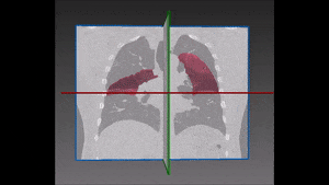
Computed tomography (CT) of the chest provides an effective and non-invasive way to study pulmonary morphology and functioning. Accurate registration of thoracic CT is in fact extremely useful to assess the clinical situation of the patient and give him or her appropriate and timely treatment. On the other hand, thoracic CT registration is also extraordinarily challenging due to the elastic nature of lung tissue deformations. Different CT lung images of the same patient suffer from local deformation, mainly due to the processes of inspiration and expiration. It is therefore necessary to stabilize the images through a transformation mapping between the different configurations of the lung, which will enable calculation of local deformation and data comparison within and (when needed) across patients.

Registration of chest from CT images

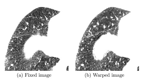
For this chest CT registration, we recommend a two-step approach which starts with an affine transformation: this is a linear mapping method that preserves points, straight lines, and planes. Its purpose in our methodology is to suggest an initial alignment, optimized in regard to translation, rotation, scaling and shearing. This alignment will be used in the following step, consisting in a deformable diffeomorphic transformation, a very valuable method for registering images collected from the same individual at different times or by different modalities such as MRI and CT. In particular, we have found this method very useful to assess pulmonary kinematics from lung CT images.
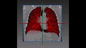
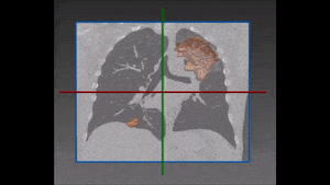
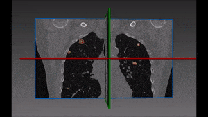
Deformation fields are evaluated in four categories: lung boundaries, fissure alignments, labeled landmarks and singularities in the deformation. Measurement data show that most lung boundaries and fissures were correctly aligned with errors close to zero. Nearly no deformation singularities were found owing to the properties of diffeomorphic transformation models.
This fully automatic method of thoracic CT registration proves itself significantly effective, independently from the source of the CT scans and regardless of the health status of the patient they were taken from. Also, its results are reliable for scans taken at any phase of the breathing cycle. Running times of each procedure of the chest CT registration are still relatively long, suggesting that future work should require finding more sophisticated ways of computation for the numerous iterations which this method necessitates.
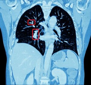
* Source: American Lung Association

Share

Share on linkedin
Share on twitter
Share on facebook

Main Field

Pulmonology

RSIP Vision’s image processing expertise in medical imaging is currently used in numerous projects, including many applications of computer vision in pulmonology. We are very proud of our contribution to lung medicine: feature detection, lung segmentation and countless other works in the pulmonary imaging field enabled our clients to give the means to physicians to save lives by giving faster and more appropriate treatment to all kind of lung diseases. You can read below about some of our breakthrough developments in Pulmonology and Bronchoscopy.

View Pulmonology

Categories

  • Pulmonology, RSIP Vision Learns

Related Content

Pulmonary embolism

Detecting Pulmonary Embolism from CT Scan

Lung vasculature segmentation

Lung tumors

Lung Tumor Segmentation

Lung Nodules Segmentation

Lung fissures

Lung Fissures Segmentation

Visible lung cancer on CT scan of chest and abdomen

Chest CT Scan Analysis with Deep Learning

Pulmonary embolism

Detecting Pulmonary Embolism from CT Scan

Lung vasculature segmentation

Lung tumors

Lung Tumor Segmentation

Lung Nodules Segmentation

Lung fissures

Lung Fissures Segmentation

Visible lung cancer on CT scan of chest and abdomen

Chest CT Scan Analysis with Deep Learning

Show all

RSIP Vision

Field-tested software solutions and custom R&D, to power your next medical products with innovative AI and image analysis capabilities.

Read more about us

Get in touch

Please fill the following form and our experts will be happy to reply to you soon

Recent News

Announcement – XPlan.ai Confirms Premier Precision in Peer-Reviewed Clinical Study of its 2D-to-3D Knee Reconstruction Solution

IBD Scoring – Clario, GI Reviewers and RSIP Vision Team Up

RSIP Neph Announces a Revolutionary Intra-op Solution for Partial Nephrectomy Surgeries

Announcement – XPlan.ai by RSIP Vision Presents Successful Preliminary Results from Clinical Study of it’s XPlan 2D-to-3D Knee Bones Reconstruction

All news
Upcoming Events
Stay informed for our next events
Subscribe to Our Magazines

Subscribe now and receive the Computer Vision News Magazine every month to your mailbox

 
Subscribe for free
Follow us
Linkedin Twitter Facebook Youtube

contact@rsipvision.com

Terms of Use

Privacy Policy

© All rights reserved to RSIP Vision 2023

Created by Shmulik

  • Our Work
    • title-1
      • Ophthalmology
      • Uncategorized
      • Ophthalmology
      • Pulmonology
      • Cardiology
      • Orthopedics
    • Title-2
      • Orthopedics
  • Success Stories
  • Insights
  • The company