Skip to content
  • Our Work
    • Fields
      • Cardiology
      • ENT
      • Gastro
      • Orthopedics
      • Ophthalmology
      • Pulmonology
      • Surgical
      • Urology
      • Other
    • Modalities
      • Endoscopy
      • Medical Segmentation
      • Microscopy
      • Ultrasound
  • Success Stories
  • Insights
    • Magazine
    • Events and Webinars
    • News
    • Blog
  • The company
    • About us
    • Careers
Menu
  • Our Work
    • Fields
      • Cardiology
      • ENT
      • Gastro
      • Orthopedics
      • Ophthalmology
      • Pulmonology
      • Surgical
      • Urology
      • Other
    • Modalities
      • Endoscopy
      • Medical Segmentation
      • Microscopy
      • Ultrasound
  • Success Stories
  • Insights
    • Magazine
    • Events and Webinars
    • News
    • Blog
  • The company
    • About us
    • Careers
Contact

Surgical

How to use visual intelligence and AI in surgical applications?

RSIP Vision brings broad and deep experience to the table for developing novel algorithmic solutions. We can help you build the advanced surgical intelligence tools to improve the efficiency, safety, surgeon training, visual enhancement, 3D navigation and more for the OR of the future. Our unique experience with surgical robotics procedures can drive your next generation solutions. We have executed successful projects involving tool & tissue tracking, registration between different coordinate systems and segmenting tools and tissues

AI and Computer Vision Technologies

From tissue segmentation to motion tracking to detection to action recognition and analytics-we’ll bring the latest state-of-the-art capabilities into your surgical applications. Our team has vast experience with a set of different modalities, such as fluoroscopic images, CT, depth images, RGB images, and heat cameras, which enables our customers to quickly introduce innovative systems into the market

2D Tool Tracking

+
2D Tool Tracking
  • Tissue tracking throughout the procedure
  • Highlight the tissue even when deformed or obscured by surgical tools
  • Improve procedural accuracy by identifying target tissue at all times

3D Tracking

+
3D Tracking
  • Use RGB or IR cameras to track objects in the Operating Room
  • Electromagnetic tracking can be used for catheter positioning
  • Implement SLAM to track endoscopes
  • Improve surgical precision by positioning Operating Room tools in the patient’s coordinate system

3D Measurements

+
3D Measurements
  • Incorporate depth perception and 3D coordinate measurements into surgical procedures
  • Accurate placement of tools relative to the patient anatomy
  • Precise measurement of pathologies
  • Improve procedural outcome and reduce risk of perforation

Pre-Op Planning

+
Pre-Op Planning
  • Choose the correct procedure before surgery starts
  • Find adequate tools that need to be used
  • Improve the accuracy and precision of surgical procedures
  • Enhance the ability to simulate and plan for potential surgical challenges
  • Reduce risk of surgical complications
  • Reduce the need for additional surgical procedures
  • Improve communication and coordination among the surgical team

Tissue Tracking

+
Tissue Tracking
  • Real-time tracking of various tissues
  • Handle deformation of soft tissues
  • Improve accuracy and precision of surgical procedures
  • Reduce tissue trauma and improved ability to preserve healthy tissue
  • Enhance the ability to monitor and respond to changes in the surgical field in real-time

3D Reconstruction

+
3D Reconstruction
  • Live and accurate 3D modeling
  • Reconstruction of anatomy
  • Reconstruction of robotic instruments
  • Internal RGB images
  • External X-ray images

3D Depth Estimation

+
3D Depth Estimation
  • Using both mono and stereo cameras
  • Improve understanding of the surgical scene
  • Improve registration of pre-operative imaging with real-time surgical video.
  • Accurate placement of tools relative to the anatomy
  • Real time response
  • Relative depth

Post-op Analysis

+
Post-op Analysis
  • Cost savings by identifying and reducing complications
  • Improved patient satisfaction and surgical outcome
  • Identify areas for future improvement in surgical techniques or protocols
  • Reduce complications and identify problematic patterns or trends

Scene Recognition

+
Scene Recognition
  • Allows for optimal tracking of surgical progress
  • Improve accuracy and precision of surgical procedures
  • Enhance ability to analyze and evaluate surgical performance
  • Increase ability to identify and preserve critical structures

Image Enhancement

+
Image Enhancement
  • AI-powered advanced denoising techniques
  • Automated image quality grading
  • Color corrections - tailored to specific modality & AI enabled
  • Context-aware enhancement based on prior anatomical knowledge

Multi-modality Registration

+
Multi-modality Registration
  • Solving orientation, resolution, FOV and dimensionality
  • Optimization-base registration
  • Classical & deep learning based technologies
  • Customized to user needs
  • 2D-to-2D, 2D-to-3D, and 3D-to-3D capabilities
  • Rigid and deformable registration
  • Support all modalities & image characteristics: CT MRI, X-ray, US and RGB

Robotics Expertise

+
Robotics Expertise
  • Facilitate the development of customized solutions for specific medical needs and requirements
  • Increase ability to design and develop devices for minimally invasive procedures
  • Provide unique expertise working with surgical robotics teams
  • Enhance ability to automate and streamline complex procedures

Case Studies

Surgical Stages Recognition

The Challenge:
  • An Industry leader wanted to automate surgical stages recognition to drive efficiencies in growing analytics pipeline
Our Approach:
  • We performed a deep literature review, providing our client with a fully updated map of technologies
  • We implemented our solution, selected to achieve best results in minimal development time
  • We then adapted this solution to multiple types of surgery
Results:
  • Our client achieved state-of-the-art stages recognition performance
  • Our solution has been integrated in our client’s software pipeline

Endoscopic Video Analysis

The challenge
  • An innovative start-up initiative in the endoscopy space wanted to quickly prove out a range of AI benefits for video analysis application
Our approach
  • The RSIP Vision R&D team, in close collaboration with in-house and client medical experts developed a multi-functional proof of concept
  • We quickly & efficiently built key capabilities including pathology detection, image segmentation, motion estimation, and image quality improvements
  • During the development process, RSIP Vision discovered novel AI use cases with strong additional business potential, enhancing our client’s value proposition
The outcome
  • Our support of this client led to a successful transaction with a large industry-leading corporation
  • We developed & delivered novel core technologies, providing a strong basis for full solution development
  • At this time, production and solution development is underway in continued partnership with RSIP Vision

Learn more

Learn more solutions and technologies from this fiel
Automated IBD Scoring

IBD Scoring – Clario, GI Reviewers and RSIP Vision Team Up

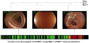
Clario, GI Reviewers and RSIP Vision Team Up to Present a New AI Solution to Advance Clinical Trials for Inflammatory Bowel Diseases Innovative, human-level AI technology will improve efficiency and consistency of Inflammatory Bowel Disease (IBD) scoring, advancing clinical trials of novel treatments for these debilitating ailments. PHILADELPHIA, PA, Copenhagen, Denmark; San Mateo, CA; Boston, MA; & Jerusalem, Israel – March 6, 2023 – Clario, a leading healthcare research and technology company that generates the richest clinical evidence for the clinical trials industry and GI Reviewers, LLC Gastroenterology consultants and

Read More

Next Generation Intra-op Navigation

During minimally invasive surgeries, surgeons are limited in their field of view, which restricts their observation of the patient’s full anatomy. This may lead to suboptimal surgical outcomes or even errors. For example, in a tumor excision procedure, healthy tissue may be injured, or worse, the surgeon may fail to optimally remove the lesion. Surgeons base the procedure on pre-op images of the area such as CT scans, which provide a snapshot of the internal tissue. Navigation systems are then utilized to fit the scan to the coordinate system of

Read More
AI in Diagnostic ERCP

Image Analysis and AI in Diagnostic ERCP

Recent developments in the field of medical image analysis and artificial intelligence (AI) are used to improve the procedural outcomes of ERCP (Endoscopic retrograde cholangiopancreatography). Here is how RSIP Vision develops AI for diagnostic ERCP. Read what we do for enabling 3D Image Reconstruction and Image Registration/Fusion. Learn how strictures detection and classification can provide the physicians with classification scoring and, sometimes, help them avoid unnecessary biopsies during ERCP.

Read More
Type 4 cholangiocarcinoma

Enhanced ERCP tumor assessment using AI

ERCP involves both endoscopy and fluoroscopy on a region with limited access. Accordingly, it poses several challenges for the gastroenterologist performing the procedure: imaging and artificial intelligence are key technologies to solve these challenges. They are used to reconstruct multiangle 2D X-ray images into a 3D image that will help the gastroenterologists in real-time navigation, reducing any complications involved in the navigation process. AI can be trained to accurately classify each tumor found by ERCP and check whether it is cancerous or not. 

Read More
ERCP Gallstones and strictures

AI in ERCP gallstones and strictures treatment

When performing ERCP for stone removal and stricture treatment, the gastroenterologist must overcome several challenges, namely navigation in the region of interest and the choice of the most fitting treatment that must be selected and implemented. AI enables to reconstructing a 3D image from multi-angle X-ray images or ultrasound slices. It is also trained to accurately classify the type of blockage that necessitated the ERCP procedure in the first place, resulting in quicker and more efficient ERCP procedures.

Read More

AI for Gastric Cancer Detection

Upper gastrointestinal cancers, including esophageal cancer and gastric cancer, are among the most common cancers worldwide. However, a lack of endoscopists with colonoscopy skills has been identified and solutions are critically needed. The development of a real-time robust detection system for colorectal neoplasms is needed to significantly reduce the risk of missed lesions during colonoscopy. 

Read More
Improved PCNL

Improved PCNL with Computer Vision

Larger stones in the kidney and proximal ureter need to be treated with PCNL (Percutaneous Nephrolithotomy), a minimally invasive urology procedure intended to remove them. It is today a very effective procedure, as it can be operated via a relatively small opening with a reduced time of hospitalization. There is also a version of it called mini-PCNL, a tubeless procedure with an even smaller port of entry, while maintaining a similar rate of efficacy. More surgeons would be able to perform PCNL if the access part were made simpler. That

Read More
The Power of AI in Robotic Surgeries

Fireside Chat – The Power of AI in Robotic Surgeries

This Fireside Chat was held on Tuesday September 26, 2023 Guest speaker: Simone Crivellaro, MD, MHA UIC, Urology Dept, Vice Chair, Lawrence S. Ross, Professor & Associate Professor UIC, Urology Robotic, Section Chief UIC, Urology at Mount Sinai Hospital, Division Chief A Fireside Chat on cutting-edge technologies behind surgical advancement, where technology seamlessly integrates with medical expertise. We discuss advanced pre-operative planning, case-specific simulations, and AI-based tools that make all these realistic, representing the future of surgery. Intra-op technologies, such as dynamic fusion of real-time ultrasound, that enhance visualization and

Read More
AI-Assisted Prostate cancer diagnosis

AI-Assisted Prostate Cancer Diagnosis

Prostate cancer is the second most prevalent cancer in men, affecting about 1 in 8 men during their lifetime. Often, patients will have an MRI scan as part of regular screening. MRI images identify regions and details such as the prostate boundary, zones and tumors, which radiologists use to calculate the Prostate Imaging Reporting and Data System (PI-RADS) score. The PI-RADS score ranges from 1 to 5 (low to high probability of clinically significant cancer) and provides the basis for diagnosis and treatment. Patients with a PI-RADS score of 3

Read More
Prostate Guidance

Intra-op Prostate Guidance by RSIP Vision

Approximately one million prostate biopsies are performed each year in the United States to diagnose and monitor prostate cancer. Doctors commonly use transrectal ultrasound (TRUS) to generate real-time images of the prostate to help guide the biopsy. To mitigate the risk of missing a lesion, physicians usually carry out multiple collections from the gland. Nonetheless, there is a risk that some tumors may go undetected. Therefore, it is common practice for the patient to have a pre-op MRI scan, which generates a more detailed image of the prostate. This provides

Read More
Soft Tissues Tracking during Brain Surgery

Soft Tissue Tracking during Brain Surgeries

Most of the tissue handled by the surgeon (or by a robotic hand) during brain surgeries is flexible: With the obvious exception of bone, which is rigid, all the structures touched during the surgery change shape and place. For instance, by changing the force applied to the brain, like when the skull is opened, the pressure on the brain changes and, as a consequence, its shape changes too. It is key for the surgeon to know where each point has moved to: if the tumor that needs to be treated

Read More
Neph - Partial Nephrectomy surgery

RSIP Neph Announces a Revolutionary Intra-op Solution for Partial Nephrectomy Surgeries

This next generation robotic surgery innovation utilizes AI-based visual intelligence to provide vital information that is overlaid and presented on the video feed in real-time during surgery  Jerusalem, Israel, Jan 26, 2023 – Today, RSIP Neph (part of the RSIP Vision group) announced its new innovative intra-op solution that utilizes advanced Computer Vision and Artificial Intelligence algorithms, to enhance the next generation of robotic surgery. This solution has been under development with the largest urology department in Israel and it will assist surgeons during the surgery itself, by providing additional

Read More
Show more

Get in touch

Please fill the following form and our experts will be happy to reply to you soon

Recent News

Announcement – XPlan.ai Confirms Premier Precision in Peer-Reviewed Clinical Study of its 2D-to-3D Knee Reconstruction Solution

IBD Scoring – Clario, GI Reviewers and RSIP Vision Team Up

RSIP Neph Announces a Revolutionary Intra-op Solution for Partial Nephrectomy Surgeries

Announcement – XPlan.ai by RSIP Vision Presents Successful Preliminary Results from Clinical Study of it’s XPlan 2D-to-3D Knee Bones Reconstruction

All news
Upcoming Events
Stay informed for our next events
Subscribe to Our Magazines

Subscribe now and receive the Computer Vision News Magazine every month to your mailbox

 
Subscribe for free
Follow us
Linkedin Twitter Facebook Youtube

contact@rsipvision.com

Terms of Use

Privacy Policy

© All rights reserved to RSIP Vision 2023

Created by Shmulik

  • Our Work
    • title-1
      • Ophthalmology
      • Uncategorized
      • Ophthalmology
      • Pulmonology
      • Cardiology
      • Orthopedics
    • Title-2
      • Orthopedics
  • Success Stories
  • Insights
  • The company