Skip to content
  • Our Work
    • Fields
      • Cardiology
      • ENT
      • Gastro
      • Orthopedics
      • Ophthalmology
      • Pulmonology
      • Surgical
      • Urology
      • Other
    • Modalities
      • Endoscopy
      • Medical Segmentation
      • Microscopy
      • Ultrasound
  • Success Stories
  • Insights
    • Magazine
    • Upcoming Events
    • Webinars
    • Meetups
    • News
    • Blog
  • The company
    • About us
    • Careers
Menu
  • Our Work
    • Fields
      • Cardiology
      • ENT
      • Gastro
      • Orthopedics
      • Ophthalmology
      • Pulmonology
      • Surgical
      • Urology
      • Other
    • Modalities
      • Endoscopy
      • Medical Segmentation
      • Microscopy
      • Ultrasound
  • Success Stories
  • Insights
    • Magazine
    • Upcoming Events
    • Webinars
    • Meetups
    • News
    • Blog
  • The company
    • About us
    • Careers
Contact

Soft Tissue Tracking during Brain Surgeries

Most of the tissue handled by the surgeon (or by a robotic hand) during brain surgeries is flexible: With the obvious exception of bone, which is rigid, all the structures touched during the surgery change shape and place. For instance, by changing the force applied to the brain, like when the skull is opened, the pressure on the brain changes and, as a consequence, its shape changes too.

Soft Tissues Tracking during Brain Surgery

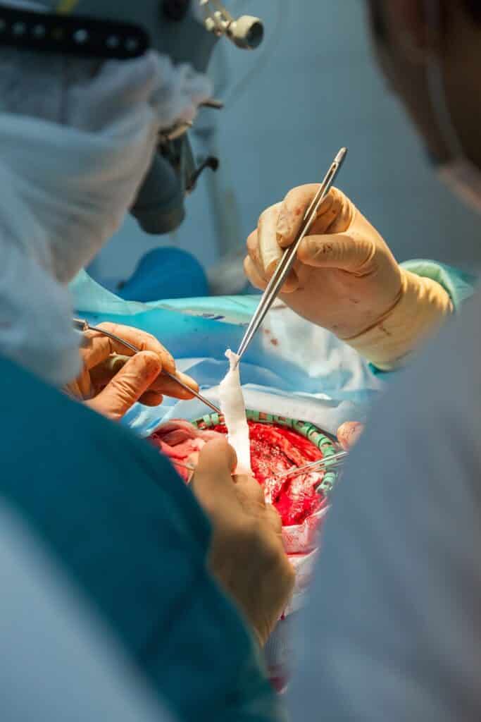
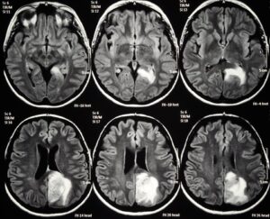
It is key for the surgeon to know where each point has moved to: if the tumor that needs to be treated was in one place, where is it now? This is a very important challenge that needs to be solved by the surgical team in real time.

Until very recently, surgeons made assumptions: for instance, they assumed that the motion is rigid. This assumption is obviously incorrect, but it was the best available hint to what was going on in the treated region. Of course, the most expert surgeons could do excellent work even with some amount of guessing. The better the human expertise involved, the better the guess – and, generally, the outcome as well.

Unfortunately, the opposite case could also be true: physicians at the beginning of their career might lack the educated guess of their seniors and, as a consequence, may bring about less positive outcomes.

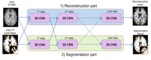
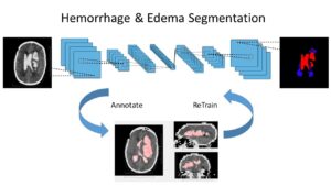
Very encouraging advances have seen the light in recent years, in particular in the field of AI-assisted image analysis; new technologies now give access to better solutions, providing a precious assistance to the surgical teams. Some of these breakthroughs are adding a whole set of possibilities to previously existing classics, like segmentation and feature tracking. In particular, advanced deep learning algorithms can process calculations much faster and offer much more reliable results; they can do that in real time, during the surgery, when supporting information is most needed.

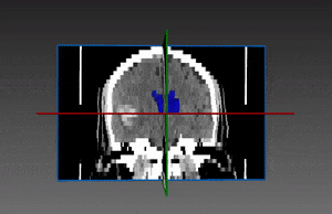
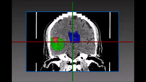
For instance, if the surgical team wants to focus on a specific region, they could start by running a segmentation of that region as a first step, and decide to track what they deem important to be tracked, choosing at will what soft tissues need to be followed. They can even decide to ignore surgical tools and remove them from the field of view as if they weren’t obstructing the eye of the surgeon. Key landmarks correspondence is done much better, making it easier to take crucial decisions regarding the physical soft tissue being treated.

These new solutions, thanks to advanced AI algorithms, are much more timely, accurate and reliable than ever before. They are more stable and, as such, more convenient, since they need fewer human corrections, many of which had some level of guessing. When old algorithms did their calculations, these were valid only for some time until the next important change in the region, after which the whole calculating process needed to be started over again. Now it is much easier to keep track even of the soft tissues in the operated region. RSIP Vision’s engineers are fully trained to develop soft tissue tracking solutions that are crucial during brain surgeries. We will be happy to discuss your project.

Share

Share on linkedin
Share on twitter
Share on facebook

Main Field

Neurology

The treatment of numerous pathologies originated in the brain is going through a revolution caused by Artificial Intelligence. In all the complexity and sophistication of the brain and regardless of the many secrets it still keeps for scientists, more and more studies are helping in the detection of brain diseases as well in their classification and the choice of a treatment: tumors, hemorrhages, traumas, strokes, exposures to radiations or chemicals, metastases, infections, genetic abnormalities and other severe conditions benefit form the progress in AI technology. Image processing of the brain, powered by deep learning techniques, is able today to give answers still unthinkable not long ago. See below some of RSIP Vision's research and projects in the area of computer vision for neurology and brain healthcare.

View Neurology

Categories

  • blog, Neurology, Robotics, RSIP Vision Learns, Surgical, Surgical

Related Content

brain ventricles

Brain Ventricles Segmentation with Deep Learning

Brain hemorrhage segmentation

Brain Hemorrhage Segmentation with Deep Learning

Joint reconstruction and segmentation

Deep Learning in Brain Imaging

Hemorrhage and Edema segmentation with Deep Learning

Intracranial Hemorrhage and Edema Segmentation

Axial cut of brain MRI

Brain Lesion Detection in MRI Images

Show all

RSIP Vision

Field-tested software solutions and custom R&D, to power your next medical products with innovative AI and image analysis capabilities.

Read more about us

Get in touch

Please fill the following form and our experts will be happy to reply to you soon

Recent News

IBD Scoring – Clario, GI Reviewers and RSIP Vision Team Up

RSIP Neph Announces a Revolutionary Intra-op Solution for Partial Nephrectomy Surgeries

Announcement – RSIP Vision Presents Successful Preliminary Results from Clinical Study of 2D-to-3D Knee Bones Reconstruction

Announcement – New Urological AI Tool for 3D Reconstruction of the Ureter

All news
Upcoming Events
Stay informed for our next events
Subscribe to Our Magazines

Subscribe now and receive the Computer Vision News Magazine every month to your mailbox

 
Subscribe for free
Follow us
Linkedin Twitter Facebook Youtube

contact@rsipvision.com

Terms of Use

Privacy Policy

© All rights reserved to RSIP Vision 2023

Created by Shmulik

  • Our Work
    • title-1
      • Ophthalmology
      • Uncategorized
      • Ophthalmology
      • Pulmonology
      • Cardiology
      • Orthopedics
    • Title-2
      • Orthopedics
  • Success Stories
  • Insights
  • The company