Skip to content
  • Our Work
    • Fields
      • Cardiology
      • ENT
      • Gastro
      • Orthopedics
      • Ophthalmology
      • Pulmonology
      • Surgical
      • Urology
      • Other
    • Modalities
      • Endoscopy
      • Medical Segmentation
      • Microscopy
      • Ultrasound
  • Success Stories
  • Insights
    • Magazine
    • Events and Webinars
    • News
    • Blog
  • The company
    • About us
    • Careers
Menu
  • Our Work
    • Fields
      • Cardiology
      • ENT
      • Gastro
      • Orthopedics
      • Ophthalmology
      • Pulmonology
      • Surgical
      • Urology
      • Other
    • Modalities
      • Endoscopy
      • Medical Segmentation
      • Microscopy
      • Ultrasound
  • Success Stories
  • Insights
    • Magazine
    • Events and Webinars
    • News
    • Blog
  • The company
    • About us
    • Careers
Contact

AI in ERCP gallstones and strictures treatment

The main challenges in ERCP

Endoscopic retrograde cholangiopancreatography (ERCP) is an intricate procedure for assessing and treating problems in the biliary ductal system. Two such problems include the presence of gallstones (cholelithiasis) and benign or malignant strictures (sclerosis, tumors or inflammation).

When performing ERCP for stone removal and stricture treatment, the gastroenterologist must overcome several challenges:

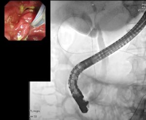
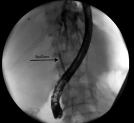
  • Navigation – navigating to the bile duct and beyond it, is a crucial step during any ERCP procedure. The delicate and complex navigation process starts in the mouth, through the esophagus, down into the stomach and through the duodenum before nearing the bile duct. Once reached, the main challenges are 1 – to enter the CBD and 2- to select the route without risk of interfering with the pancreas. Then, contrast agent is introduced in order to image and locate the blockage. The gastroenterologist then needs to gain full access to the cause of the blockage and treat it accordingly.
  • Treatment selection – once the blockage has been reached, the gastroenterologist needs to select and implement the most fitting treatment. If the blockage is caused by cholelithiasis, typically a lithotripter, basket or Fogarty balloon will be used, and if the cause is a stricture, usually a fitting stent will be inserted.
ERCP Gallstones and strictures
Common bile duct stones as seen in CT

Imaging and AI in ERCP

The following advanced  image analysis and artificial intelligence technologies can help tackle the challenges involved in treating stones and strictures, making the procedure easier, quicker and safer.

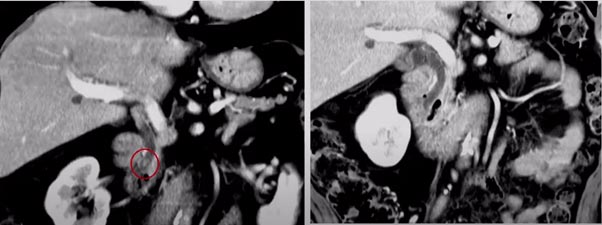
  • 3D reconstruction – reconstructing a 3D image from multi-angle X-ray images or ultrasound slices can help the gastroenterologist reach any blockage. Using a 3D reconstructed image during this interventional procedure will help not only in navigating to the bile duct and beyond it, but also in assessing the blockage and selecting the best course of treatment, since it will provide an improved understanding of the blockage in 3 dimensions, and allow accurate measurements of the needed areas, for quantitative diagnosis and treatment planning.
  • Blockage classification – accurately imaging the cause of the blockage is key in treating it. Several imaging modalities are used for ERCP procedures, among them are computerized tomography (CT), X-ray and magnetic resonance cholangiopancreatography (MRCP). Deep learning and machine learning methods can be trained to accurately classify the blockage using features regarding geometry and texture as either cholelithiasis or strictures (either benign or malignant).

RSIP Vision and ERCP

RSIP Vision has extensive knowledge and experience in developing technologies that assist surgeons in interventional procedures using 3D reconstructed images. This hands-on experience is highly applicable to ERCP procedures, in which navigation is essential and complications are unfortunately common.

Additionally, RSIP Vision’s proven record in image analysis and AI can help classify the type of blockage that necessitated the ERCP procedure in the first place. By using deep learning and machine learning methods to accurately determine the cause of the blockage, the gastroenterologists will be better informed and prepared for treatment.

Using techniques of image analysis and AI in ERCP will result in quicker and more efficient procedures, with fewer complications, and better overall treatment for the patient.

Share

Share on linkedin
Share on twitter
Share on facebook

Main Field

Gastro

Artificial Intelligence is more and more used for detection, diagnosis and treatment of gastrointestinal diseases. Image analysis techniques are very helpful to power classical endoscopic tools and to helps classify all kind of items, including lesions, colorectal polyps, tumors, bleedings, occlusions and more. Today's most modern AI for GI software powers surgical robots developed to effectively intervene in the most dire gastroenterology procedures and cases.

View Gastro

Categories

  • Gastro, RSIP Vision Learns

Related Content

Automated IBD Scoring

IBD Scoring – Clario, GI Reviewers and RSIP Vision Team Up

AI in Diagnostic ERCP

Image Analysis and AI in Diagnostic ERCP

Type 4 cholangiocarcinoma

Enhanced ERCP tumor assessment using AI

AI for Gastric Cancer Detection

Gastrointestinal lesion detection with machine learning

Show all

RSIP Vision

Field-tested software solutions and custom R&D, to power your next medical products with innovative AI and image analysis capabilities.

Read more about us

Get in touch

Please fill the following form and our experts will be happy to reply to you soon

Recent News

Announcement – XPlan.ai Confirms Premier Precision in Peer-Reviewed Clinical Study of its 2D-to-3D Knee Reconstruction Solution

IBD Scoring – Clario, GI Reviewers and RSIP Vision Team Up

RSIP Neph Announces a Revolutionary Intra-op Solution for Partial Nephrectomy Surgeries

Announcement – XPlan.ai by RSIP Vision Presents Successful Preliminary Results from Clinical Study of it’s XPlan 2D-to-3D Knee Bones Reconstruction

All news
Upcoming Events
Stay informed for our next events
Subscribe to Our Magazines

Subscribe now and receive the Computer Vision News Magazine every month to your mailbox

 
Subscribe for free
Follow us
Linkedin Twitter Facebook Youtube

contact@rsipvision.com

Terms of Use

Privacy Policy

© All rights reserved to RSIP Vision 2023

Created by Shmulik

  • Our Work
    • title-1
      • Ophthalmology
      • Uncategorized
      • Ophthalmology
      • Pulmonology
      • Cardiology
      • Orthopedics
    • Title-2
      • Orthopedics
  • Success Stories
  • Insights
  • The company