Skip to content
  • Our Work
    • Fields
      • Cardiology
      • ENT
      • Gastro
      • Orthopedics
      • Ophthalmology
      • Pulmonology
      • Surgical
      • Urology
      • Other
    • Modalities
      • Endoscopy
      • Medical Segmentation
      • Microscopy
      • Ultrasound
  • Success Stories
  • Insights
    • Magazine
    • Events and Webinars
    • News
    • Blog
  • The company
    • About us
    • Careers
Menu
  • Our Work
    • Fields
      • Cardiology
      • ENT
      • Gastro
      • Orthopedics
      • Ophthalmology
      • Pulmonology
      • Surgical
      • Urology
      • Other
    • Modalities
      • Endoscopy
      • Medical Segmentation
      • Microscopy
      • Ultrasound
  • Success Stories
  • Insights
    • Magazine
    • Events and Webinars
    • News
    • Blog
  • The company
    • About us
    • Careers
Contact

Meibomian gland dysfunction detection

Meibomian gland dysfunction is often seen as an early stage of dry eye syndrome. Indeed, Meibomian glands play a significant role in tears production by contributing lipids to the superficial tear film. Pathologies which occur in meibomian glands limit the secretion of oil, causing poor lubrication (lubrification) of the eye and severe irritation.
Tear film formation and meibomian glands
Meibomian glands and tear film formation
Three imaging procedures exist to detect this pathology: the first method uses infrared meibography; the second uses laser confocal meibography; the third uses Optical Coherence Tomographic meibography.

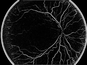
Infrared meibography is an imaging technique producing excellent quality images of meibomian glands, but not sufficient to provide information at the microscopic level. For this purpose, other technologies have been proposed, like the laser confocal microscopy, which uses laser scans to acquire very clear and precise images of the microscopic structures of the meibomian glands. However, this type of meibography is slightly more invasive, since it requires eyelid eversion: to decrease pain and facilitate procedure, topical anesthesia is applied before performing alignment of the lens against the posterior aspect of the everted eyelid.

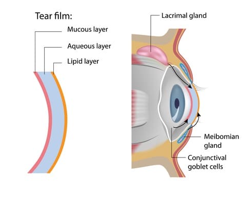
Evaluation of Meibomian Gland Dysfunction

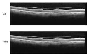
The advanced way to evaluate meibomian gland dysfunction is by using highly accurate OCT, which delivers a 3D tomography of the glands. It may display features and measurements which are crucial to understand the specific problem of each gland.

The image analysis of the gland is very complex, because what is seen is not one organ with one contour: it looks like a grape-like structure, which in the normal eye is composed of many small clustered parts. In the abnormal eye, the observer will see differences in the topography of the gland which need to be explained. Filtering techniques for noise reduction and thresholding allow to perform a correct segmentation of the gland. Fourier Transform is the main algorithmic technique enabling us to decompose the image and perform the image processing operations used to detect and describe the presence of meibomian gland dysfunction: this and other tools help to accurately assess the regularity of the gland and of its structure. RSIP Vision has decades-long experience in these advanced imaging procedures. Share with us your work and your projects!

Share

Share on linkedin
Share on twitter
Share on facebook

Main Field

Ophthalmology

RSIP Vision has developed countless projects in the field of ophthalmology for its clients. Computer vision with sophisticated ophthalmic imaging, measurement techniques and AI can yield better results: great precision, accurate diagnosis and best interventional treatment for many pathologies.

View Ophthalmology

Categories

  • Ophthalmology, RSIP Vision Learns

Related Content

Super-Resolution in OCT images

Super-Resolution in OCT images

Zoom-in-Net

Deep Learning in Ophthalmology

Classification and Segmentation of Dendritic cells

Classification and Segmentation of Dendritic Cells

Alzheimer's Disease - AD

Degenerative Diseases Detection in the Eye

Temporary pediatric strabismus in newborn baby

Image processing for pediatric strabismus

ROP - Vessel tortuosity in Retinopathy of Prematurity

ROP: Retinopathy of Prematurity

Super-Resolution in OCT images

Super-Resolution in OCT images

Zoom-in-Net

Deep Learning in Ophthalmology

Classification and Segmentation of Dendritic cells

Classification and Segmentation of Dendritic Cells

Alzheimer's Disease - AD

Degenerative Diseases Detection in the Eye

Temporary pediatric strabismus in newborn baby

Image processing for pediatric strabismus

ROP - Vessel tortuosity in Retinopathy of Prematurity

ROP: Retinopathy of Prematurity

Show all

RSIP Vision

Field-tested software solutions and custom R&D, to power your next medical products with innovative AI and image analysis capabilities.

Read more about us

Get in touch

Please fill the following form and our experts will be happy to reply to you soon

Recent News

Announcement – XPlan.ai Confirms Premier Precision in Peer-Reviewed Clinical Study of its 2D-to-3D Knee Reconstruction Solution

IBD Scoring – Clario, GI Reviewers and RSIP Vision Team Up

RSIP Neph Announces a Revolutionary Intra-op Solution for Partial Nephrectomy Surgeries

Announcement – XPlan.ai by RSIP Vision Presents Successful Preliminary Results from Clinical Study of it’s XPlan 2D-to-3D Knee Bones Reconstruction

All news
Upcoming Events
Stay informed for our next events
Subscribe to Our Magazines

Subscribe now and receive the Computer Vision News Magazine every month to your mailbox

 
Subscribe for free
Follow us
Linkedin Twitter Facebook Youtube

contact@rsipvision.com

Terms of Use

Privacy Policy

© All rights reserved to RSIP Vision 2023

Created by Shmulik

  • Our Work
    • title-1
      • Ophthalmology
      • Uncategorized
      • Ophthalmology
      • Pulmonology
      • Cardiology
      • Orthopedics
    • Title-2
      • Orthopedics
  • Success Stories
  • Insights
  • The company