Skip to content
  • Our Work
    • Fields
      • Cardiology
      • ENT
      • Gastro
      • Orthopedics
      • Ophthalmology
      • Pulmonology
      • Surgical
      • Urology
      • Other
    • Modalities
      • Endoscopy
      • Medical Segmentation
      • Microscopy
      • Ultrasound
  • Success Stories
  • Insights
    • Magazine
    • Events and Webinars
    • News
    • Blog
  • The company
    • About us
    • Careers
Menu
  • Our Work
    • Fields
      • Cardiology
      • ENT
      • Gastro
      • Orthopedics
      • Ophthalmology
      • Pulmonology
      • Surgical
      • Urology
      • Other
    • Modalities
      • Endoscopy
      • Medical Segmentation
      • Microscopy
      • Ultrasound
  • Success Stories
  • Insights
    • Magazine
    • Events and Webinars
    • News
    • Blog
  • The company
    • About us
    • Careers
Contact

Super-Resolution in OCT images

OCT images are frequently used to demonstrate the retina and understand the different pathologies and decide the diagnostics and help with procedure planning.

In medical imaging, especially in radiology, deep learning has been used in recent years to gain super-resolution. On one hand, it is possible to scan faster and more efficiently at lower resolutions. On the other hand, quality scans provide added benefits. Super-resolution is a class of techniques in image processing that enhance the resolution of an imaging system, making images sharper and clearer. By improving the quality of OCT images, super-resolution can potentially enhance our ability to detect and analyze pathological changes, thus facilitating more accurate and early diagnosis of various eye conditions.

Super-Resolution in OCT images
The figure above shows the prediction image as compared to the ground truth, where prediction was restored from an image with a resolution reduced by a factor of 2. Pay attention to the fact that we are always dealing with images in a given size. What superresolution or upsampling gives us is “more slices”, meaning the distance between slices will be lower because we have predicted the slices between them.

This is why we use super-resolution also in OCT images and get better resolution or – alternatively – gain scanning speed to produce images with the quality needed for the diagnostics. RSIP Vision has already done that: developing super-resolution for OCT images. It can also be useful in real-time scans, when high quality images are needed in real-time.

Several steps are indicated for the typical process, as follows.

It is crucial to select a training dataset which includes distinctive pathologies in the right proportion. Otherwise, the algorithm might be trained only for healthy eyes and would offer no clinical value. Each pathology needs to be represented in the dataset, and this for several reasons: first, to train the system also on that pathology; also, to declare to the regulatory authorities that the new algorithm won’t miss any meaningful diagnostics. From the point of view of the compliance officer – he needs to assure that no pathology is missed due to the process. The way to do it is to run on a list of typical pathologies and validate it one by one.

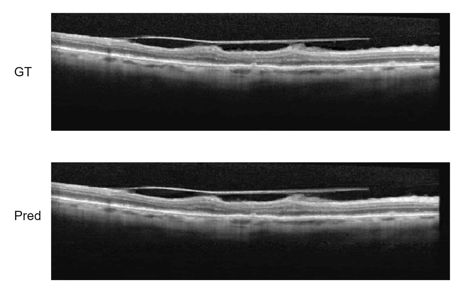
The training itself of the convolutional neural network will take advantage of several architectures for improving the resolution: in particular, Super-resolution CNN (SR-CNN): a model is trained on patches of OCT scan at low resolution, where higher resolution scan is known. In this manner, we go from small patches to more detailed patches by inferring what the model knows about expected retinal anatomy. We have proven that, using a minimal dataset of specific OCT scans, we were able to generalize and have a super-resolutional model with good results..

It is also very sensitive to check the loss function and adjust it to the specific case of OCT images, which abound in stain-like textures stemming from speckles that are not meaningful. The algorithm engineer has to adjust the loss function accordingly.

RSIP Vision has developed a very precise super-resolution algorithm for OCT. Contact us to check how we can use it for your AI project in ophthalmology.


Share

Share on linkedin
Share on twitter
Share on facebook

Main Field

Ophthalmology

RSIP Vision has developed countless projects in the field of ophthalmology for its clients. Computer vision with sophisticated ophthalmic imaging, measurement techniques and AI can yield better results: great precision, accurate diagnosis and best interventional treatment for many pathologies.

View Ophthalmology

Categories

  • blog, Ophthalmology, RSIP Vision Learns

Related Content

Zoom-in-Net

Deep Learning in Ophthalmology

Classification and Segmentation of Dendritic cells

Classification and Segmentation of Dendritic Cells

Alzheimer's Disease - AD

Degenerative Diseases Detection in the Eye

Temporary pediatric strabismus in newborn baby

Image processing for pediatric strabismus

ROP - Vessel tortuosity in Retinopathy of Prematurity

ROP: Retinopathy of Prematurity

Eyelid Drooping - MRD1 and MRD2

Eyelid Drooping – Blepharoptosis

Show all

RSIP Vision

Field-tested software solutions and custom R&D, to power your next medical products with innovative AI and image analysis capabilities.

Read more about us

Get in touch

Please fill the following form and our experts will be happy to reply to you soon

Recent News

Announcement – XPlan.ai Confirms Premier Precision in Peer-Reviewed Clinical Study of its 2D-to-3D Knee Reconstruction Solution

IBD Scoring – Clario, GI Reviewers and RSIP Vision Team Up

RSIP Neph Announces a Revolutionary Intra-op Solution for Partial Nephrectomy Surgeries

Announcement – XPlan.ai by RSIP Vision Presents Successful Preliminary Results from Clinical Study of it’s XPlan 2D-to-3D Knee Bones Reconstruction

All news
Upcoming Events
Stay informed for our next events
Subscribe to Our Magazines

Subscribe now and receive the Computer Vision News Magazine every month to your mailbox

 
Subscribe for free
Follow us
Linkedin Twitter Facebook Youtube

contact@rsipvision.com

Terms of Use

Privacy Policy

© All rights reserved to RSIP Vision 2023

Created by Shmulik

  • Our Work
    • title-1
      • Ophthalmology
      • Uncategorized
      • Ophthalmology
      • Pulmonology
      • Cardiology
      • Orthopedics
    • Title-2
      • Orthopedics
  • Success Stories
  • Insights
  • The company