Skip to content
  • Our Work
    • Fields
      • Cardiology
      • ENT
      • Gastro
      • Orthopedics
      • Ophthalmology
      • Pulmonology
      • Surgical
      • Urology
      • Other
    • Modalities
      • Endoscopy
      • Medical Segmentation
      • Microscopy
      • Ultrasound
  • Success Stories
  • Insights
    • Magazine
    • Upcoming Events
    • Webinars
    • Meetups
    • News
    • Blog
  • The company
    • About us
    • Careers
Menu
  • Our Work
    • Fields
      • Cardiology
      • ENT
      • Gastro
      • Orthopedics
      • Ophthalmology
      • Pulmonology
      • Surgical
      • Urology
      • Other
    • Modalities
      • Endoscopy
      • Medical Segmentation
      • Microscopy
      • Ultrasound
  • Success Stories
  • Insights
    • Magazine
    • Upcoming Events
    • Webinars
    • Meetups
    • News
    • Blog
  • The company
    • About us
    • Careers
Contact

Brain Ventricles Segmentation with Deep Learning

The brain ventricular system is in charge of cerebrospinal fluid (CSF) production, which is essential for its normal function. In addition to providing physical protection to the brain tissue, it provides the nervous system with nutrients and removes waste among other functions. The ventricular system consists of four ventricles: two lateral ventricles in each hemisphere, and third and fourth ventricles, all connected by several narrow foramina. Due to its great importance and complexity, early diagnosis and treatment of ventricular system pathologies is crucial. For example, outflow blockage may cause ventricular enlargement due to increased pressure. Computed tomography (CT) imaging of the brain has become a leading diagnostic tool due to its high availability and quick image generation, which is useful in emergency room settings such as stroke or traumatic brain injury (TBI). CT imaging can supply the physicians with crucial information regarding presence of hemorrhage, ischemia, tumors, hydrocephalus, and other pathologies.

brain ventricles

One of the most problematic consequences of TBI, stroke or brain tumor is increased intracranial pressure (ICP), which is a direct indicator of poor outcome when rising to dangerous levels. Mostly, increased ICP is due to intracranial hematoma or cerebral edema, and can lead to a decreased blood supply to brain tissue due to displacement of brain structures called mass effect, brain herniation, and hydrocephalus. Hence, early diagnosis and treatment of increased ICP is essential for minimizing mortality.

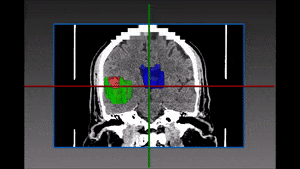
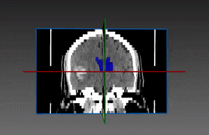
There are several features in imaging that might suggest that the patient is suffering from increased ICP or is at risk for developing it. Two of them are – shifting of the midline and increased size of the lateral ventricles. Presence of these features could indicate the need for invasive treatments such as decompressive craniotomy or placement of a catheter within one of the lateral ventricles called external ventricular drain (EVD) , in order to drain cerebrospinal fluid (CSF) and relieve pressure from brain tissue.

Brain ventricles segmentation with AI

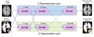
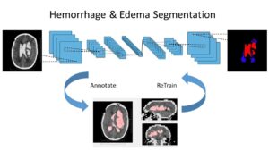
It is therefore key to accurately segment lateral ventricles and the midline. The main challenges resides in the following: hemorrhage or tumors which are external to the ventricle (like in ICH) may modify its shape due to external pressure; also, when hemorrhage are within the ventricle (IVH), blood can fill the volume of the ventricle rendering the segmentation too difficult for traditional medical imaging techniques. RSIP Vision has developed a cutting edge deep neural network to perform a very accurate brain ventricles segmentation through advanced Artificial Intelligence techniques. Talk to an RSIP Vision engineer now.

Share

Share on linkedin
Share on twitter
Share on facebook

Main Field

Neurology

The treatment of numerous pathologies originated in the brain is going through a revolution caused by Artificial Intelligence. In all the complexity and sophistication of the brain and regardless of the many secrets it still keeps for scientists, more and more studies are helping in the detection of brain diseases as well in their classification and the choice of a treatment: tumors, hemorrhages, traumas, strokes, exposures to radiations or chemicals, metastases, infections, genetic abnormalities and other severe conditions benefit form the progress in AI technology. Image processing of the brain, powered by deep learning techniques, is able today to give answers still unthinkable not long ago. See below some of RSIP Vision's research and projects in the area of computer vision for neurology and brain healthcare.

View Neurology

Categories

  • Neurology, RSIP Vision Learns

Related Content

Soft Tissues Tracking during Brain Surgery

Soft Tissue Tracking during Brain Surgeries

Brain hemorrhage segmentation

Brain Hemorrhage Segmentation with Deep Learning

Joint reconstruction and segmentation

Deep Learning in Brain Imaging

Hemorrhage and Edema segmentation with Deep Learning

Intracranial Hemorrhage and Edema Segmentation

Axial cut of brain MRI

Brain Lesion Detection in MRI Images

Show all

RSIP Vision

Field-tested software solutions and custom R&D, to power your next medical products with innovative AI and image analysis capabilities.

Read more about us

Get in touch

Please fill the following form and our experts will be happy to reply to you soon

Recent News

IBD Scoring – Clario, GI Reviewers and RSIP Vision Team Up

RSIP Neph Announces a Revolutionary Intra-op Solution for Partial Nephrectomy Surgeries

Announcement – RSIP Vision Presents Successful Preliminary Results from Clinical Study of 2D-to-3D Knee Bones Reconstruction

Announcement – New Urological AI Tool for 3D Reconstruction of the Ureter

All news
Upcoming Events
Stay informed for our next events
Subscribe to Our Magazines

Subscribe now and receive the Computer Vision News Magazine every month to your mailbox

 
Subscribe for free
Follow us
Linkedin Twitter Facebook Youtube

contact@rsipvision.com

Terms of Use

Privacy Policy

© All rights reserved to RSIP Vision 2023

Created by Shmulik

  • Our Work
    • title-1
      • Ophthalmology
      • Uncategorized
      • Ophthalmology
      • Pulmonology
      • Cardiology
      • Orthopedics
    • Title-2
      • Orthopedics
  • Success Stories
  • Insights
  • The company