Skip to content
  • Our Work
    • Fields
      • Cardiology
      • ENT
      • Gastro
      • Orthopedics
      • Ophthalmology
      • Pulmonology
      • Surgical
      • Urology
      • Other
    • Modalities
      • Endoscopy
      • Medical Segmentation
      • Microscopy
      • Ultrasound
  • Success Stories
  • Insights
    • Magazine
    • Upcoming Events
    • Webinars
    • Meetups
    • News
    • Blog
  • The company
    • About us
    • Careers
Menu
  • Our Work
    • Fields
      • Cardiology
      • ENT
      • Gastro
      • Orthopedics
      • Ophthalmology
      • Pulmonology
      • Surgical
      • Urology
      • Other
    • Modalities
      • Endoscopy
      • Medical Segmentation
      • Microscopy
      • Ultrasound
  • Success Stories
  • Insights
    • Magazine
    • Upcoming Events
    • Webinars
    • Meetups
    • News
    • Blog
  • The company
    • About us
    • Careers
Contact

Brain Hemorrhage Segmentation with Deep Learning

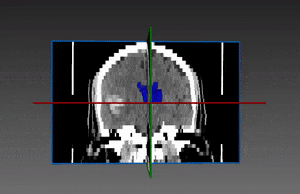
Intracranial hemorrhage is defined as bleeding within the cranium. Bleeding can occur within the brain parenchyma itself, giving way to what is called intra-axial bleed or intracerebral hemorrhage. On the other hand, bleeding outside the brain tissue is referred to as extra-axial bleed. Subtypes of extra axial bleed include epidural, subdural and subarachnoid hemorrhages.

Brain hemorrhage segmentation
Brain hemorrhage segmentation – in red. Ventricles are in blue and edema in green.

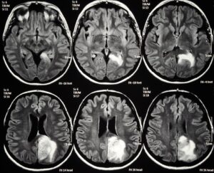
The intracranial hemorrhage might cause a Phenomenon called “mass effect”, meaning that the hemorrhage causes areas of brain tissue or brain structures surrounding it to be compressed and injured, due to elevated pressure in the restricted space within the skull. This could result, together with the edema around it, in elevated intracranial pressure (ICP). Elevated ICP is correlated with poor outcome, and may result in intubation, mechanical ventilation, multiple complications; if left untreated, it can be fatal. Thus, early diagnosis, monitoring and treatment are essential.

Two types of intracranial bleeding that cause hemorrhagic stroke are intracerebral hemorrhage and subarachnoid hemorrhage. Intracerebral or subarachnoid bleed can be accompanied by intraventricular hemorrhage (IVH), which is a bleeding into the brain’s ventricular system. IVH might occur after head trauma or as a result of hemorrhagic stroke. In this case as well, the volume of the bleeding is a predictive factor of prognosis and a valid method of evaluation is needed.

Automated brain hemorrhage segmentation

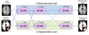
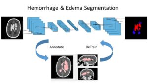
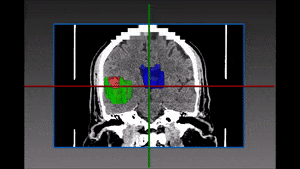
RSIP Vision’s image analysis algorithm module based on deep learning can rapidly estimate the hemorrhage volume and measure the edematous area around it for both ICH and IVH. Some cases of brain hemorrhage may require ICP monitoring and drainage via intra-ventricular catheter. Our image processing algorithms produce a 3D model of the ventricular system, which can ultimately be useful in guidance of the neurosurgeon during ventriculostomy.

SAH (subarachnoid hemorrhage) segmentation is challenging due to very variable and detailed shape, together with low contrast on CT. RSIP Vision used a large quantity of annotated data to feed a deep learning neural network able to segment all kinds of hemorrhages and accurately measure their volume. Cutting-edge Convolutional Neural Networks (CNN) architecture has been instrumental to achieve accurate results. Call our engineers to make sure that this module is what you are looking for.

Share

Share on linkedin
Share on twitter
Share on facebook

Main Field

Neurology

The treatment of numerous pathologies originated in the brain is going through a revolution caused by Artificial Intelligence. In all the complexity and sophistication of the brain and regardless of the many secrets it still keeps for scientists, more and more studies are helping in the detection of brain diseases as well in their classification and the choice of a treatment: tumors, hemorrhages, traumas, strokes, exposures to radiations or chemicals, metastases, infections, genetic abnormalities and other severe conditions benefit form the progress in AI technology. Image processing of the brain, powered by deep learning techniques, is able today to give answers still unthinkable not long ago. See below some of RSIP Vision's research and projects in the area of computer vision for neurology and brain healthcare.

View Neurology

Categories

  • Neurology, RSIP Vision Learns

Related Content

Soft Tissues Tracking during Brain Surgery

Soft Tissue Tracking during Brain Surgeries

brain ventricles

Brain Ventricles Segmentation with Deep Learning

Joint reconstruction and segmentation

Deep Learning in Brain Imaging

Hemorrhage and Edema segmentation with Deep Learning

Intracranial Hemorrhage and Edema Segmentation

Axial cut of brain MRI

Brain Lesion Detection in MRI Images

Show all

RSIP Vision

Field-tested software solutions and custom R&D, to power your next medical products with innovative AI and image analysis capabilities.

Read more about us

Get in touch

Please fill the following form and our experts will be happy to reply to you soon

Recent News

IBD Scoring – Clario, GI Reviewers and RSIP Vision Team Up

RSIP Neph Announces a Revolutionary Intra-op Solution for Partial Nephrectomy Surgeries

Announcement – RSIP Vision Presents Successful Preliminary Results from Clinical Study of 2D-to-3D Knee Bones Reconstruction

Announcement – New Urological AI Tool for 3D Reconstruction of the Ureter

All news
Upcoming Events
Stay informed for our next events
Subscribe to Our Magazines

Subscribe now and receive the Computer Vision News Magazine every month to your mailbox

 
Subscribe for free
Follow us
Linkedin Twitter Facebook Youtube

contact@rsipvision.com

Terms of Use

Privacy Policy

© All rights reserved to RSIP Vision 2023

Created by Shmulik

  • Our Work
    • title-1
      • Ophthalmology
      • Uncategorized
      • Ophthalmology
      • Pulmonology
      • Cardiology
      • Orthopedics
    • Title-2
      • Orthopedics
  • Success Stories
  • Insights
  • The company