Brain surgeries are very complex: they need to be extremely accurate, since you want to spare healthy tissue; planning is very thorough, as the surgeon looks in multi-imaging modalities – for instance, where the tumor is located, what needs to be removed, and what is the best path; execution of the plan needs to be very precise, avoiding any damage to any surrounding structure, in a patient that is not always fully anesthetized.
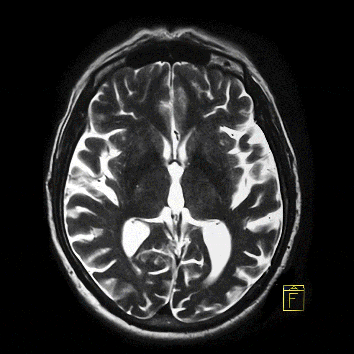
It is also a long and gruesome process, in which mistakes that sometimes happen have dire consequences. You do not want to leave a person paralyzed, because the surgeon slightly diverges from the optimal path. At a recent keynote at the Hamlyn Symposium, Professor Alexandra Golby spoke from her experience as a neurosurgeon, developing AI systems for brain surgery. She took the problem one step further. There are pathways within the brain: highways of neurons that should be avoided if possible. Under specific modalities and MRI sequences, these highways can be seen. This information is very precious for the surgeon to avoid them.
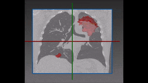
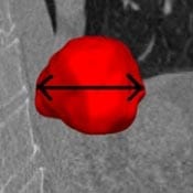
How can Artificial Intelligence help? All levels of the specific surgery process can be assisted by a specific AI tool. Let’s start with the planning: standard procedure calls for MRI protocols to visualize the brain and the pathology in the brain, helping to plan a path. Where AI becomes handy is by doing an automatic segmentation of the brain, of its surrounding substructures, and of the skull, so that we can visually see everything. The tumor is also segmented, shown in 3d adjacent to all the anatomical structures . Once this model is ready, the surgeon can devise the ideal path for accessing the tumor. This is not always a straight line; it might be a curved path. A good 3d model allows the surgeon to identify the entry point and the path that will the least harm surrounding tissues of the brain. This can be taken one step further, by devising curved tools that best follow a curved path.
There is also a need for multimodalities registration, since the tumor may look different in different MRI protocols or CT scan. Once they are registered, each modality contributes vital information and all can be seen as one for better assessment. Additionally, by correctly segmenting the tumor, it becomes possible to remove it accurately, sparing any tissue that should not be removed. This is specifically important for brain surgery: during tumor removal, the surgeon always takes a safety buffer around the tumor to verify no cancer cells remain. In the brain this buffer should be kept minimal, as brain tissue preservation is of high importance.
What tasks does AI perform in order to do it? Precise segmentation of the tumor, precise segmentation of all the surrounding structures, accurate registration of all different modalities, as different MRI protocols result in different images. The radiologists know which one is providing the better assessment or whether a synthesis of multiple protocols should be accepted to make sure that the entire tumor is perfectly segmented. These things being done, with a good planning software the surgeon can visualize everything and prepare a successful procedure.
Does AI play a role also during the procedure itself? The answer is yes, and it is a very crucial one. It’s not enough to have a plan, it must also be executed adequately. But more challenges await the surgical team: the table can move, the patient can move on the table, brain parts can move once the skull is opened – all this needs to be accounted for in real time. Intra-operative imaging modalities are already used: X-rays, ultrasound and even MRI is used. All these modalities need to be registered with the pre-op scan to best follow the original plan. This needs to be extremely precise and this is why AI should be employed. It needs to accurately compensate for the motion of the patient, and for the patient’s brain within the skull so that the registration is flawless at all times and the surgeon can follow the plan.
AI plays a role also in the follow-up steps. Traditionally, MRI scans are used for post-op assessment of tumor removal. Any subsequent growth or any remaining pathology must be noted as soon as possible. As differences between different scans and protocols can be subtle, AI can perform this comparison accurately and efficiently. This comparison is key for any clinical decision making, and AI can assist in making the best decision.
The AI algorithms that can perform all this at real-time need to be very refined and sophisticated: much work needs to be done by the developers to make sure that these AI algorithms for neurosurgery are clinical-grade. All these tasks, however, are not co-dependent: they do not need to be developed at once. Each task requires a solution on its own, and together they can create the ideal neurosurgery.