Skip to content
  • Our Work
    • Fields
      • Cardiology
      • ENT
      • Gastro
      • Orthopedics
      • Ophthalmology
      • Pulmonology
      • Surgical
      • Urology
      • Other
    • Modalities
      • Endoscopy
      • Medical Segmentation
      • Microscopy
      • Ultrasound
  • Success Stories
  • Insights
    • Magazine
    • Upcoming Events
    • Webinars
    • Meetups
    • News
    • Blog
  • The company
    • About us
    • Careers
Menu
  • Our Work
    • Fields
      • Cardiology
      • ENT
      • Gastro
      • Orthopedics
      • Ophthalmology
      • Pulmonology
      • Surgical
      • Urology
      • Other
    • Modalities
      • Endoscopy
      • Medical Segmentation
      • Microscopy
      • Ultrasound
  • Success Stories
  • Insights
    • Magazine
    • Upcoming Events
    • Webinars
    • Meetups
    • News
    • Blog
  • The company
    • About us
    • Careers
Contact

Registration of CT and X-ray

Registration is a phase of orthopedic surgeries in which a visualization system is called to support the surgeon. This system provides real time view of the surgery environment and the information from the CT imaging.
3D CT Scan
3D CT Scan
Two different steps of registration are executed here: tracking system registration and C-Arm X-ray to CT registration.

Registration between tracking system and CT scan

When the tracking system and the CT scan successively combines and no misalignment is found, a registration between them is achieved. In this way, every movement and orientation of the surgery tool is easily translated into a combined view, offering the surgeon in the operation room a very powerful visual aid to perform the intended procedure.

2D C-Arm scan
2D C-Arm X-ray scan

Registration of CT and X-ray images

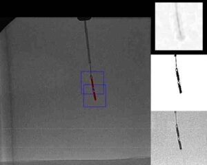
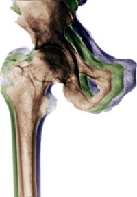
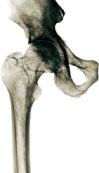
This step correlates the bones scanned, during pre-operation’s CT scan with bones scanned in-op (during the operation) via C-Arm (X-Ray). The algorithm gets a set of C-Scan 2D images (from the X-Ray) and a 3D scan (from CT). Using an optimization process, the 2D scans are accurately correlated to the 3D scan. The algorithm uses the pixel size obtained from the calibration model to make sure the bones are matched (object-wise) even when scans resolutions (2D to 3D) differ.

2D C-Arm X-ray scan is registered to 3D CT scan

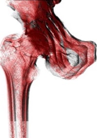
The yield transformation Registration of CT and X-ray here shows the location of each pixel of the C-Arm scans in the CT 3D scan. In this way, Registration of CT and X-ray is achieved.

This is the third in a series of five articles about Orthopedic Navigation. Read the next article about Navigation system tools in Orthopedic Surgeries. You can also read about RSIP Vision’s other works in Orthopedic Surgery.

Share

Share on linkedin
Share on twitter
Share on facebook

Main Field

General Medical

View General Medical

Categories

  • General Medical, RSIP Vision Learns

Related Content

MRI - AI-Assisted Brain Surgery

AI-Assisted Brain Surgery

Automated RECIST score

Detection and Tracking of Tumors

Pharma - RECIST score

Automated RECIST Measurement

Navigation Systems for Surgery

Catheter Navigation System with RL

U-Net network architecture

Deep Learning in Medical Imaging

Magnetic Resonance Imaging Software - Catheter

Magnetic Resonance Imaging software

Show all

RSIP Vision

Field-tested software solutions and custom R&D, to power your next medical products with innovative AI and image analysis capabilities.

Read more about us

Get in touch

Please fill the following form and our experts will be happy to reply to you soon

Recent News

IBD Scoring – Clario, GI Reviewers and RSIP Vision Team Up

RSIP Neph Announces a Revolutionary Intra-op Solution for Partial Nephrectomy Surgeries

Announcement – RSIP Vision Presents Successful Preliminary Results from Clinical Study of 2D-to-3D Knee Bones Reconstruction

Announcement – New Urological AI Tool for 3D Reconstruction of the Ureter

All news
Upcoming Events
Stay informed for our next events
Subscribe to Our Magazines

Subscribe now and receive the Computer Vision News Magazine every month to your mailbox

 
Subscribe for free
Follow us
Linkedin Twitter Facebook Youtube

contact@rsipvision.com

Terms of Use

Privacy Policy

© All rights reserved to RSIP Vision 2023

Created by Shmulik

  • Our Work
    • title-1
      • Ophthalmology
      • Uncategorized
      • Ophthalmology
      • Pulmonology
      • Cardiology
      • Orthopedics
    • Title-2
      • Orthopedics
  • Success Stories
  • Insights
  • The company