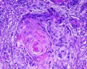
Multiplex IF analysis allows for real-time relational data between cells in tissue. This is an advantage over the gold standard brightfield IHC, which looks at a single marker at a time. When using brightfield IHC for multiple markers, sequential sections of the tissue are needed which does not allow for cell-cell relational data as you are looking at different cells between each section. What Multiplex does is that it allows a nice view of the immune landscape of the tissue so that you can study the interaction between multiple cells within that same tissue.
Still, several challenges are on the way of the researcher: it is more challenging on the wet-lab component, due to the higher complexity of performing multiple stains. It is also challenging on the analysis side, for several reasons:
• When you are dealing with fluorescence, you have background auto-fluorescence, of which you must get rid to be able to do work.
• There is always a risk of cross-bleed of the wavelength between the channels which can lead to false positive results. It is necessary to ensure that what you are seeing is a real signal and not a leak from another channel.
• Also, some of these markers are challenging to separate because the cells might be very close together, or even overlapping. It is essential to accurately separate these cells so that you can correctly phenotype the cells.
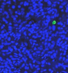
• There are also cell types that may or may not contain a visible nucleus which makes detection a challenge. On the nuclear side you also have cells of different shape, size and intensity making conventional segmentation approaches very challenging.
Multiplex IF analysis with Deep Learning
RSIP Vision’s solution to the Multiplex task is a fully automated deep learning approach: we are not looking for hard thresholds to segment the nuclei; we prefer feeding exactly the nuclei that we see into our deep learning model and creating the network that will detect all of them. Our neural network does really well in detecting the nuclei (regardless of the shape, the intensity, the size) and also at separating them. This makes our technology ideal for pharma companies doing research and translational work, for cancer centers investigating exploratory results from clinical trial data, or anyone trying to understand either the mechanism of action of specific drugs or the immediate status of a patient undergoing a clinical trial. This is the perfect companion solution in immune-oncology, where it is crucial to understand the current immune landscape of a patient.
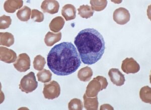
 Microscopy
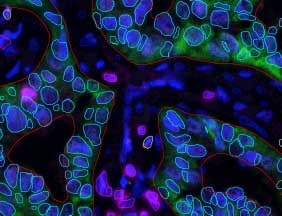