Skip to content
  • Our Work
    • Fields
      • Cardiology
      • ENT
      • Gastro
      • Orthopedics
      • Ophthalmology
      • Pulmonology
      • Surgical
      • Urology
      • Other
    • Modalities
      • Endoscopy
      • Medical Segmentation
      • Microscopy
      • Ultrasound
  • Success Stories
  • Insights
    • Magazine
    • Upcoming Events
    • Webinars
    • Meetups
    • News
    • Blog
  • The company
    • About us
    • Careers
Menu
  • Our Work
    • Fields
      • Cardiology
      • ENT
      • Gastro
      • Orthopedics
      • Ophthalmology
      • Pulmonology
      • Surgical
      • Urology
      • Other
    • Modalities
      • Endoscopy
      • Medical Segmentation
      • Microscopy
      • Ultrasound
  • Success Stories
  • Insights
    • Magazine
    • Upcoming Events
    • Webinars
    • Meetups
    • News
    • Blog
  • The company
    • About us
    • Careers
Contact

Cell Classification software

One ml of human blood contains roughly 5 million red blood cells. This huge quantity is only a fraction of what is found in one ml of blood, which contains roughly 60% fluid (plasma) and the remaining white cells, red blood cells and platelets. The composition of blood is examined routinely in hospitals: abnormalities in blood composition discloses potential diseases and indicate the response to treatment. For example, the presence of immature blast (white) cells in the circulation might indicate the development of leukemia, or myelodysplastic syndrome.

white blood cell classification

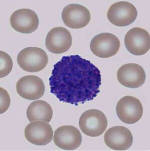
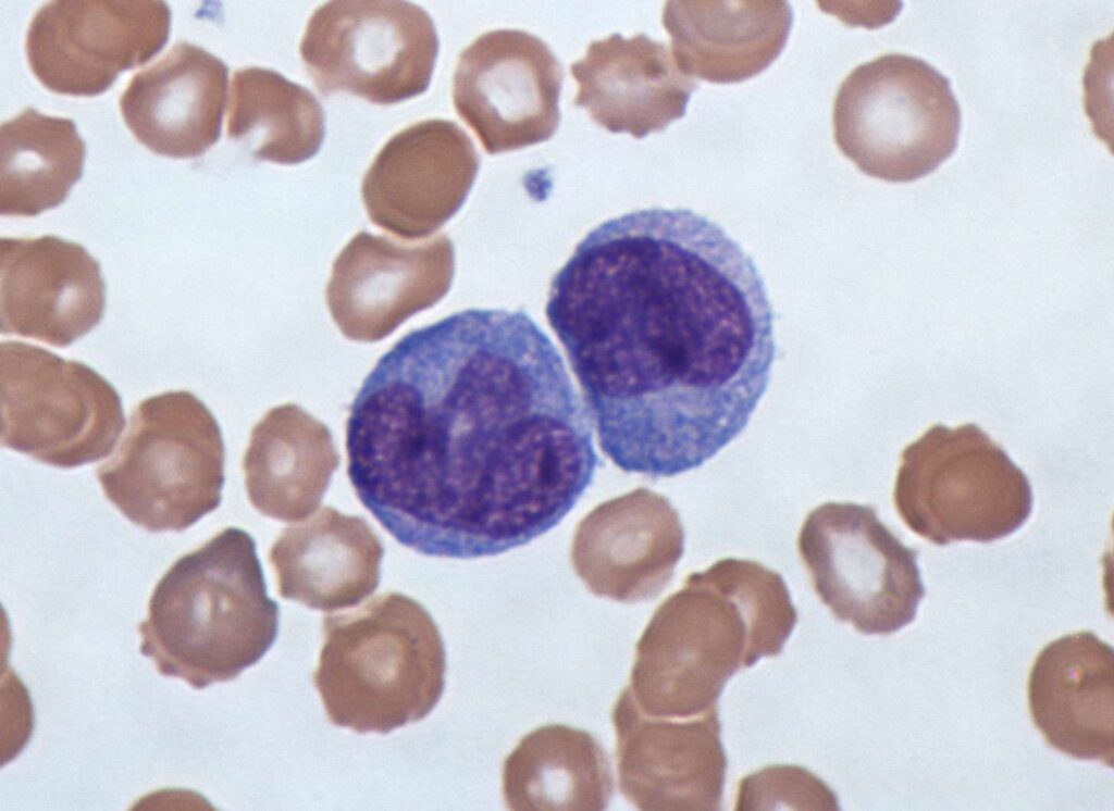
A scientific identification of the blood composition is performed by chemical means, by which antibodies for specific cell types serve as indicators for their presence and concentration. However, pathologies are many times suggested by abnormalities found in cell morphologies (enlarged nuclei, smaller overall size, etc.). Such morphological changes are usually examined in tissue biopsy by microscopic inspection for solid tissues. In the case of blood cells, morphological abnormalities need to be examined manually under the microscope in a drop of blood. However, this process is largely inefficient.
.

Automatic cell classification software

Automatic means to inspect and recognize blood cells can be achieved by computer vision and machine learning techniques. A cell classification software, built with algorithms dedicated to this process, identifies cells based on their morphological features. These features are extracted from an image of cells and fed to a machine learning classifier to obtain the final classification. By these means, blood count can be made quickly and efficiently. When blood is drawn from patients, it can be led to pass through a narrow hose, allowing the passage of only few cells at a time. A camera mounted above the hose records the blood as it flows and transmits the images to an image processing module for the extraction of features in individual cells.
.

RSIP Vision’s cell classification software

RSIP Vision has constructed a software package, allowing the recognition and count of cells based on their morphology. Machine learning algorithms were trained in collaboration with experienced hematologists, to produce a recognition rate above 97%, identifying and counting basophils, eosinophils, lymphocytes, monocytes and neutrophils.  Our algorithm far exceeds the speed at which a human can classify cells, and the accuracy rates obtained place our algorithm as the state-of-the-art in its domain.

Detection of abnormalities in terms of cell number or morphology provides means to construct an alert system for the test and pass the result to further expert examination. Our cell classification software produces detailed, efficient and effective classification of a large number of cells at a time, enabling faster cancer detection.

We’ve been developing image processing software for the biomedical industry for many years. You are welcome to contact our experts to discuss how we can help you meet your R&D challenges!
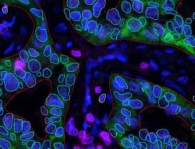
Share

Share on linkedin
Share on twitter
Share on facebook

Main Field

Microscopy

View Microscopy

Categories

  • Microscopy, RSIP Vision Learns

Related Content

Multiplex

Multiplex IF Analysis

CTCs - Circulating Tumor Cells

Circulating Tumor Cells (CTCs)

Pharma - Tissue Analysis

Tissue Analysis with AI

Pharma - Dendritic cell

Detection and Segmentation of Dendritic Cells

Cell Classification - pattern recognition classification techniques

Biomedical image processing and algorithms

Automated diagnostic reading of HIV

Show all

RSIP Vision

Field-tested software solutions and custom R&D, to power your next medical products with innovative AI and image analysis capabilities.

Read more about us

Get in touch

Please fill the following form and our experts will be happy to reply to you soon

Recent News

IBD Scoring – Clario, GI Reviewers and RSIP Vision Team Up

RSIP Neph Announces a Revolutionary Intra-op Solution for Partial Nephrectomy Surgeries

Announcement – RSIP Vision Presents Successful Preliminary Results from Clinical Study of 2D-to-3D Knee Bones Reconstruction

Announcement – New Urological AI Tool for 3D Reconstruction of the Ureter

All news
Upcoming Events
Stay informed for our next events
Subscribe to Our Magazines

Subscribe now and receive the Computer Vision News Magazine every month to your mailbox

 
Subscribe for free
Follow us
Linkedin Twitter Facebook Youtube

contact@rsipvision.com

Terms of Use

Privacy Policy

© All rights reserved to RSIP Vision 2023

Created by Shmulik

  • Our Work
    • title-1
      • Ophthalmology
      • Uncategorized
      • Ophthalmology
      • Pulmonology
      • Cardiology
      • Orthopedics
    • Title-2
      • Orthopedics
  • Success Stories
  • Insights
  • The company