Skip to content
  • Our Work
    • Fields
      • Cardiology
      • ENT
      • Gastro
      • Orthopedics
      • Ophthalmology
      • Pulmonology
      • Surgical
      • Urology
      • Other
    • Modalities
      • Endoscopy
      • Medical Segmentation
      • Microscopy
      • Ultrasound
  • Success Stories
  • Insights
    • Magazine
    • Events and Webinars
    • News
    • Blog
  • The company
    • About us
    • Careers
Menu
  • Our Work
    • Fields
      • Cardiology
      • ENT
      • Gastro
      • Orthopedics
      • Ophthalmology
      • Pulmonology
      • Surgical
      • Urology
      • Other
    • Modalities
      • Endoscopy
      • Medical Segmentation
      • Microscopy
      • Ultrasound
  • Success Stories
  • Insights
    • Magazine
    • Events and Webinars
    • News
    • Blog
  • The company
    • About us
    • Careers
Contact

Image Analysis and AI in Diagnostic ERCP

Endoscopic retrograde cholangiopancreatography (ERCP) is an imaging modality used for diagnosis and treatment of pathologies in the pancreatico-biliary system. Recent developments in the field of medical image analysis and artificial intelligence (AI) can be used to improve ERCP procedural outcomes. Here is how we use AI in Diagnostic ERCP.

3D Image Reconstruction for Video-Feed and X-Ray

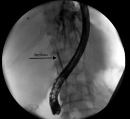
The first navigational challenge of ERCP is inserting the guidewire into the Common Bile Duct (CBD) through the Major Papilla, while avoiding the pancreatic duct. This is done using the live-video image from the endoscopic camera, and X-ray verification using dye injections.

Threading a wire through a narrow opening using 2D images can be difficult. 3D reconstruction of the endoscope or the X-ray images, using advanced image analysis algorithms, adds the depth dimension to the process, increases the real 3D structure perception which contributes to shorten the procedure time, and reduce bowel perforations.

AI in Diagnostic ERCP


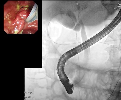
Image Registration/Fusion for ERCP

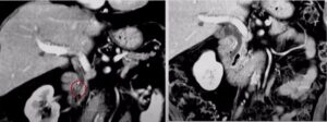
  • Baseline Scan Registration: Often a CT or MRI scan of the patient is done prior to the ERCP. Both scans provide 3-dimensional data with high resolution. Registration/Fusion of the endoscopy and/or X-ray data with the pre-op baseline scan will result in a clearer image of the strictures shape and location. Having a more decisive image for ERCP may shorten the procedure and reduce redundant dye injections, resulting in fewer cases of pancreatitis.
  • Video and X-Ray Registration: It is challenging to decipher a pathology from a single image. Advanced image analysis, utilizing C-Arm positioning and endoscope tracking, allows registration of the video and the X-ray images. This allows examination of suspected areas using 2 imaging modalities, with certainty that the same region-of-interest is displayed on both, an additional verification step needed for best treatment selection.

Stricture Classification

X-ray imaging during ERCP provides the ability to detect the strictures within the biliary ducts, but not the source and types of these strictures (gallstone, scar, or tumor) that needs to be decided by the physician or by a biopsy. Using ERCP procedures carefully annotated data, deep-learning algorithms can be trained to classify these constrained regions of the bile ducts accurately, provide the physician with classification scoring and, in some cases, avoid unnecessary biopsies during ERCP.

AI in Diagnostic ERCP by RSIP Vision

ERCP can benefit tremendously from the advancements in the field of image analysis and AI. Currently it utilizes two imaging modalities, without the additional value of processing the images and fusing the data from them. RSIP Vision has extensive development experience with medical imaging, including endoscopy and X-ray data, and can assist in optimizing this procedure. Our vast understanding of the ERCP field, combined with our experience and clinically proven technologies deployed in the market, gives us the needed edge to develop your custom next generation solution, gaining improved capabilities and speeding time-to-market.

Share

Share on linkedin
Share on twitter
Share on facebook

Main Field

Gastro

Artificial Intelligence is more and more used for detection, diagnosis and treatment of gastrointestinal diseases. Image analysis techniques are very helpful to power classical endoscopic tools and to helps classify all kind of items, including lesions, colorectal polyps, tumors, bleedings, occlusions and more. Today's most modern AI for GI software powers surgical robots developed to effectively intervene in the most dire gastroenterology procedures and cases.

View Gastro

Categories

  • Gastro, RSIP Vision Learns

Related Content

Automated IBD Scoring

IBD Scoring – Clario, GI Reviewers and RSIP Vision Team Up

Type 4 cholangiocarcinoma

Enhanced ERCP tumor assessment using AI

ERCP Gallstones and strictures

AI in ERCP gallstones and strictures treatment

AI for Gastric Cancer Detection

Gastrointestinal lesion detection with machine learning

Show all

RSIP Vision

Field-tested software solutions and custom R&D, to power your next medical products with innovative AI and image analysis capabilities.

Read more about us

Get in touch

Please fill the following form and our experts will be happy to reply to you soon

Recent News

Announcement – XPlan.ai Confirms Premier Precision in Peer-Reviewed Clinical Study of its 2D-to-3D Knee Reconstruction Solution

IBD Scoring – Clario, GI Reviewers and RSIP Vision Team Up

RSIP Neph Announces a Revolutionary Intra-op Solution for Partial Nephrectomy Surgeries

Announcement – XPlan.ai by RSIP Vision Presents Successful Preliminary Results from Clinical Study of it’s XPlan 2D-to-3D Knee Bones Reconstruction

All news
Upcoming Events
Stay informed for our next events
Subscribe to Our Magazines

Subscribe now and receive the Computer Vision News Magazine every month to your mailbox

 
Subscribe for free
Follow us
Linkedin Twitter Facebook Youtube

contact@rsipvision.com

Terms of Use

Privacy Policy

© All rights reserved to RSIP Vision 2023

Created by Shmulik

  • Our Work
    • title-1
      • Ophthalmology
      • Uncategorized
      • Ophthalmology
      • Pulmonology
      • Cardiology
      • Orthopedics
    • Title-2
      • Orthopedics
  • Success Stories
  • Insights
  • The company