Skip to content
  • Our Work
    • Fields
      • Cardiology
      • ENT
      • Gastro
      • Orthopedics
      • Ophthalmology
      • Pulmonology
      • Surgical
      • Urology
      • Other
    • Modalities
      • Endoscopy
      • Medical Segmentation
      • Microscopy
      • Ultrasound
  • Success Stories
  • Insights
    • Magazine
    • Events and Webinars
    • News
    • Blog
  • The company
    • About us
    • Careers
Menu
  • Our Work
    • Fields
      • Cardiology
      • ENT
      • Gastro
      • Orthopedics
      • Ophthalmology
      • Pulmonology
      • Surgical
      • Urology
      • Other
    • Modalities
      • Endoscopy
      • Medical Segmentation
      • Microscopy
      • Ultrasound
  • Success Stories
  • Insights
    • Magazine
    • Events and Webinars
    • News
    • Blog
  • The company
    • About us
    • Careers
Contact

Implementing AI to Improve PI-RADS Scoring

Prostate Cancer and PI-RADS scoring

Prostate cancer is the most common male cancer in the USA. When diagnosed early, mortality rates are very low, therefore, most men over the age of 60 years undergo screening for prostate cancer that includes PSA levels and digital rectal examination. Common practice dictates that when Prostate-specific antigen (PSA) levels are high, or when the patient is in a higher risk group, a prostate MRI scan is performed. This scan is read by a radiologist in search of lesions.

To set an objective diagnosis for prostate lesions in MRI, the PI-RADS (Prostate Imaging–Reporting and Data System) score was developed. It accounts for the lesions’ intensity, restriction, size, and shape, and provides a standardized score for reporting among physicians. Whenever a prostate MRI scan is conducted, the radiologist carefully examines it and determines a PI-RADS score for the patient. This process is highly subjective and time-consuming.

Prostate Tumor Segmentation
Segmentation of a tumor (blue), transitional zone (green) and peripheral zone (yellow), which are used to determine PI-RADS score together with other characteristics

PI-RADS scoring

The score ranges from 1 to 5. In PI-RADS scores 1 and 2, no further follow-up is needed unless there is a change in the clinical examination or the PSA levels. PI-RADS 3 is equivocal, for some lesions the radiologist will recommend either follow-up or biopsy. PI-RADS 4 and 5 mandate biopsy as they infer a high risk of cancer.

When follow-up is recommended, it is termed active surveillance or watchful waiting – continuous imaging and biopsies, depending on the medical recommendation. When the followed-up lesion develops into carcinoma, the surveillance is discontinued, and the urologist actively treats the cancer (surgery, chemotherapy, radiotherapy or combination therapy). In patients who are high-risk for surgery (old, debilitated, serious background diseases, etc.), even a PI-RADS score of 4-5 may lead to continuous follow-up.

In active surveillance/watchful waiting, a prostate MRI scan is performed and compared to previous scans. Any dynamic change (positive or negative) in the lesion characteristics needs to be documented and measured, as it may be the trigger to stop active surveillance and proceed to active treatment. This leads to high volumes of prostate scans which need to be analyzed by radiologists – quickly and accurately.

Artificial Intelligence in prostate MRI

Currently, the precision of the radiological follow-up is highly dependent on the physician’s experience and the scan conditions. One of the best ways to ensure objectiveness is by implementing AI and deep learning into the practice.

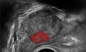
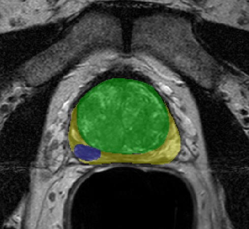
AI and computer vision algorithms are capable of segmenting, detecting, and comparing structural and visual changes in the prostate, specifically the volume, intensity and restriction of the lesions. These changes can be recorded and presented to the radiologist for review prior to diagnosis, providing robust tools for an accurate comparison.

Other Similar Use-Cases

Women at risk of breast cancer (BRCA carriers, family history) often undergo annual breast-MRI exams as a surveillance tool. Similar to prostate cancer, these scans can be automatically assessed and compared, providing an accurate map for differences between scans. By applying the same technology of the PI-RADS assistant to breast MRI, we can get a BI-RADS assistant.

RSIP Vision’s Solutions

RSIP Vision has a long, proven track record of AI and deep-learning solutions for multiple medical devices. Our team of clinicians and engineers can tailor a solution for PI-RADS scoring assistance for any device or platform. Contact us today to speed your device production and time-to-market!

Share

Share on linkedin
Share on twitter
Share on facebook

Main Field

Urology

Urology is one of the many medical fields that are being revolutionized by Artificial Intelligence. Prostate, bladder, urethra, kidneys and more key organs are already benefitting from impressive breakthroughs brought about by new technologies. AI is now able to contribute to more accurate and faster detection, segmentation, classification and diagnose for many urology disfunctions and diseases, including cancer, BPH and urolithiasis. Thanks to the latest generation of AI and computer vision techniques (deep learning in particular), AI provides now a precious support to the medical team in the operating room as well as during all stages of treatment, drastically improving the quality of therapy and reducing recovery time. You can find below some of RSIP Vision's projects and pioneering research in the field of AI for urology.

View Urology

Categories

  • blog, RSIP Vision Learns, Urology

Related Content

Improved PCNL

Improved PCNL with Computer Vision

AI-Assisted Prostate cancer diagnosis

AI-Assisted Prostate Cancer Diagnosis

Prostate Guidance

Intra-op Prostate Guidance by RSIP Vision

Neph - Partial Nephrectomy surgery

RSIP Neph Announces a Revolutionary Intra-op Solution for Partial Nephrectomy Surgeries

Benign Prostatic Hyperplasia BPH

AI for Benign Prostatic Hyperplasia BPH

Stone tracking - urology

Stone tracking during kidney stone removal

Show all

RSIP Vision

Field-tested software solutions and custom R&D, to power your next medical products with innovative AI and image analysis capabilities.

Read more about us

Get in touch

Please fill the following form and our experts will be happy to reply to you soon

Recent News

Announcement – XPlan.ai Confirms Premier Precision in Peer-Reviewed Clinical Study of its 2D-to-3D Knee Reconstruction Solution

IBD Scoring – Clario, GI Reviewers and RSIP Vision Team Up

RSIP Neph Announces a Revolutionary Intra-op Solution for Partial Nephrectomy Surgeries

Announcement – XPlan.ai by RSIP Vision Presents Successful Preliminary Results from Clinical Study of it’s XPlan 2D-to-3D Knee Bones Reconstruction

All news
Upcoming Events
Stay informed for our next events
Subscribe to Our Magazines

Subscribe now and receive the Computer Vision News Magazine every month to your mailbox

 
Subscribe for free
Follow us
Linkedin Twitter Facebook Youtube

contact@rsipvision.com

Terms of Use

Privacy Policy

© All rights reserved to RSIP Vision 2023

Created by Shmulik

  • Our Work
    • title-1
      • Ophthalmology
      • Uncategorized
      • Ophthalmology
      • Pulmonology
      • Cardiology
      • Orthopedics
    • Title-2
      • Orthopedics
  • Success Stories
  • Insights
  • The company