This article was first published on Computer Vision News of June 2022.
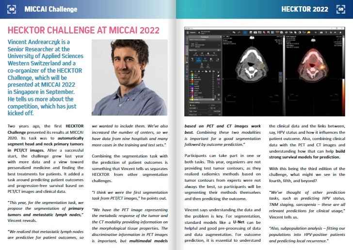
Vincent Andrearczyk is a Senior Researcher at the University of Applied Sciences Western Switzerland and a co-organizer of the HECKTOR Challenge, which will be presented at MICCAI 2022 in Singapore in September. He tells us more about the competition, which has just kicked off.
Two years ago, the first HECKTOR Challenge presented its results at MICCAI 2020. Its task was to automatically segment head and neck primary tumors in PET/CT images. After a successful start, the challenge grew last year with more data and a view toward personalized medicine and finding the best treatments for patients. It added a task around predicting patient outcomes and progression-free survival based on PET/CT images and clinical data.
“This year, for the segmentation task, we propose the segmentation of primary tumors and metastatic lymph nodes,” Vincent reveals.
“We realized that metastatic lymph nodes are predictive for patient outcomes, so we wanted to include them. We’ve also increased the number of centers, so we have data from nine hospitals and many more cases in the training and test sets.”
Combining the segmentation task with the prediction of patient outcomes is something that Vincent tells us separates HECKTOR from other segmentation challenges.
“I think we were the first segmentation task from PET/CT images,” he points out.
“We have the PET image representing the metabolic response of the tumor and the CT modality providing information on the morphological tissue properties. The discriminative information in PET images is important, but multimodal models based on PET and CT images work best. Combining these two modalities is important for a good segmentation followed by outcome prediction.”
Participants can take part in one or both tasks. This year, organizers are not providing test tumor contours, as they realized radiomics methods based on tumor contours from experts were not always the best, so participants will be segmenting their methods themselves and then predicting the outcome.
Vincent says understanding the data and the problem is key. For segmentation, standard models like a U-Net can be helpful and good pre-processing of data and data augmentation. For outcome prediction, it is essential to understand the clinical data and the links between, say, HPV status and how it influences the patient outcome. Also, combining clinical data with the PET and CT images and understanding how that can help build strong survival models for prediction.
With this being the third edition of the challenge, what might we see in the fourth, fifth, and beyond?
“We’ve thought of other prediction tasks, such as predicting HPV status, TNM staging, sarcopenia – these are all relevant predictions for clinical usage,” Vincent tells us.
“Also, subpopulation analysis – fitting our populations into HPV-positive patients and predicting local recurrence.”
Its consortium of organizers represents the interdisciplinarity of this challenge. Together, they discussed its design and analysis and how to develop guidelines for annotations. The first task seeks algorithmic contributions, but the second is about making decisions on the outcomes and performance metrics that are most meaningful for the clinic in head and neck cancer treatment, ultimately to help clinicians find the best treatment for patients.
Although the challenge is taking up a great deal of Vincent’s time now, particularly this last sprint before releasing the data, his day job focuses on deep learning and radiomics problems for radiology, including segmentation and predicting outcomes for different cancers.
“I also work on the interpretability of the models, which is very important for clinical acceptance,” he says.
“The end goal is always clinical interpretability, which is another next step for HECKTOR. Evaluating the interpretability and uncertainty of the models is very important if we want to reach clinical development.”
Keep reading the Medical Imaging News section of our magazine.