Advance navigation techniques in orthopedic surgery allow physicians to construct 3D models of patients’ anatomical parts. Such models are especially useful for surgery planning and guidance during operation. Three-dimensional model overlay on images acquired from various modalities facilitated inspection and measurements and should be routinely incorporated into the surgery pipeline.
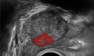
Three-dimensional models are obtained by matching (or interpolating) a surface between an acquired set of data points. Modalities for acquiring points and surface data include costly CT and MRI scans. Alternatively, intra-operative scans such as ultrasound or fluoroscopic scan can be performed with much reduced costs, health hazards and manual effort. A set of surface points marked by the technician or physician forms an initial sparse set for anatomical surface reconstruction.
Reconstruction of a surface from a sparse set of points is a challenging task from the algorithmic point of view. Sparse set of points and the high variability of possible structures makes the problem essentially ill-posed. Oftentimes shape primitives and prior information is needed to make an initial surface converge accurately to the anatomical structure based on only few landmark points. Orthopedic surgery requires high reconstruction accuracy, stability and robustness to noise, as well as reproducibility.
Incorporating prior information into the anatomical surface reconstruction process can be done using the point distribution model. This model uses a set of training points to learn possible shape variation after limiting the shape parameter space using PCR (Principal Component Analysis). Prediction of the shape based on landmark points is performed in a low dimensional shape parameter space. This approach allows one to obtain a predicted mean shape aligned to the data. Furthermore, registration can be performed using the parameters of the rigid transformation.
Following registration, a second stage of surface deformation can be applied on the mean shape, based on landmark points. Surface deformation is done with the goal of finding shape parameters which minimizes a distance measure between the surface and the landmark points while satisfying shape deformation constraints. Regularization of the function for minimization is often required in order to achieve a stable deformation convergence process.
With the incorporation of more landmark points, anatomical shape reconstruction in orthopedic surgery will improve. Allowing the process to accept new information and converge stably to a more refined shape can be achieved by the method outlined above. Intra-operative imaging accompanied by the creation of instantaneous 3D models to overlay on images is a valuable tool, which is applicable not only to orthopedic surgeries, but also to many non-deformable shape reconstruction procedures in medical imaging like in dentistry.
Shape reconstruction from sparse visual data is well within the realm of computer vision, statistics, mathematics and engineering. These qualities and expertise have been cultivated at RSIP Vision for more than two decades. Our company’s engineers are expert in computer vision, image processing and machine learning, with a long record of success in surface reconstruction for both deformable and non-deformable anatomical structure. Read our other articles about computer vision in orthopedic surgery.