Skip to content
  • Our Work
    • Fields
      • Cardiology
      • ENT
      • Gastro
      • Orthopedics
      • Ophthalmology
      • Pulmonology
      • Surgical
      • Urology
      • Other
    • Modalities
      • Endoscopy
      • Medical Segmentation
      • Microscopy
      • Ultrasound
  • Success Stories
  • Insights
    • Magazine
    • Events and Webinars
    • News
    • Blog
  • The company
    • About us
    • Careers
Menu
  • Our Work
    • Fields
      • Cardiology
      • ENT
      • Gastro
      • Orthopedics
      • Ophthalmology
      • Pulmonology
      • Surgical
      • Urology
      • Other
    • Modalities
      • Endoscopy
      • Medical Segmentation
      • Microscopy
      • Ultrasound
  • Success Stories
  • Insights
    • Magazine
    • Events and Webinars
    • News
    • Blog
  • The company
    • About us
    • Careers
Contact

AI-Assisted Prostate Cancer Diagnosis

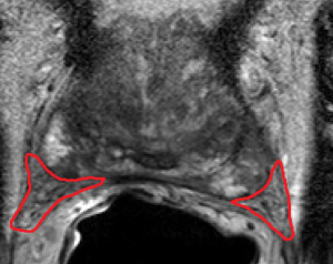
Prostate cancer is the second most prevalent cancer in men, affecting about 1 in 8 men during their lifetime. Often, patients will have an MRI scan as part of regular screening. MRI images identify regions and details such as the prostate boundary, zones and tumors, which radiologists use to calculate the Prostate Imaging Reporting and Data System (PI-RADS) score. The PI-RADS score ranges from 1 to 5 (low to high probability of clinically significant cancer) and provides the basis for diagnosis and treatment. Patients with a PI-RADS score of 3 or more usually undergo a prostate biopsy to detect suspected cancer.

AI-Assisted Prostate cancer diagnosis

Calculation of the PI-RADS score may be influenced by external factors such as experience, training or fatigue of the radiologist, which introduces variability into the process. Artificial Intelligence has the potential to serve as a valuable aid in accurately and robustly calculating the PI-RADS score, as AI algorithms output numerical values, which increase diagnosis objectivity and repeatability. For example, gradients and color distributions can be quantified to analyze the tumor and determine the homogeneity of the tissue. The data obtained from AI algorithms assists physicians in calculating the PI-RADS score, leading to a more appropriate course of action. These advances in determining crucial details such as the size, location and state of the cancer allow physicians to choose the most suitable treatment for the patient.

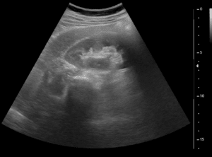
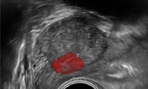
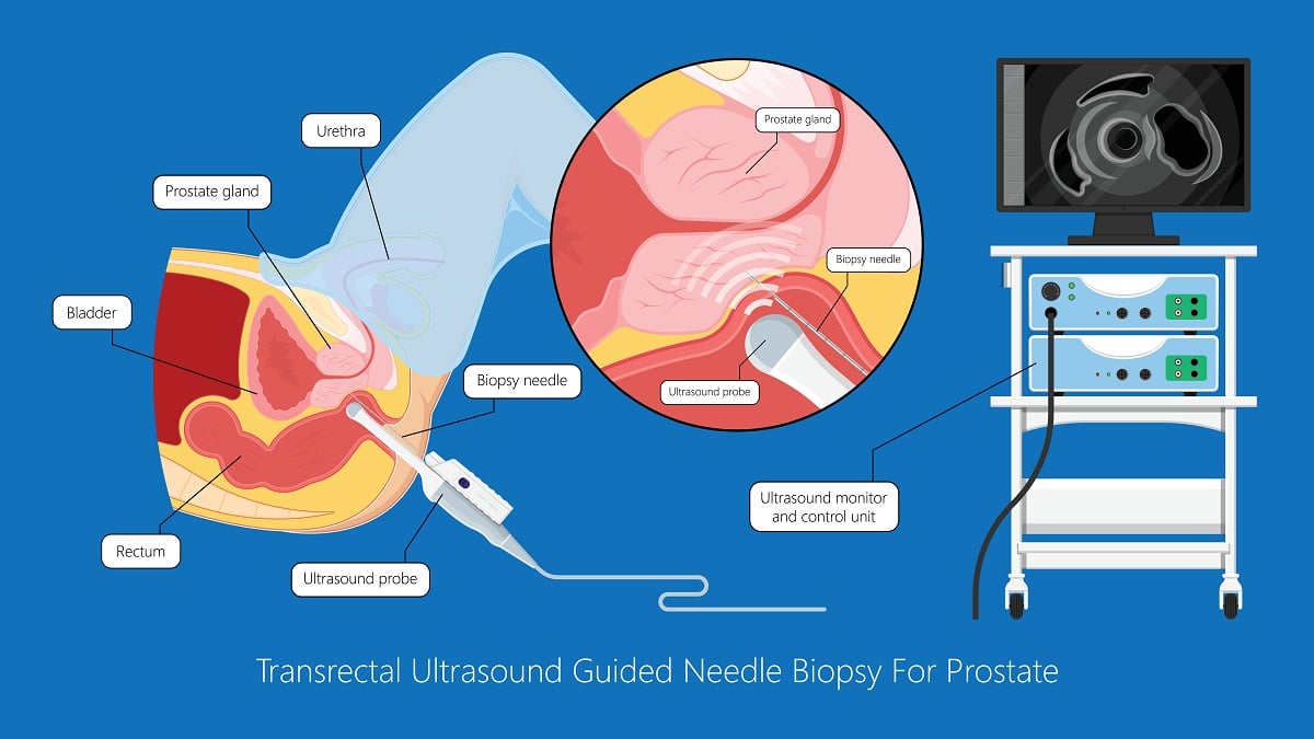
MRI-generated images of the prostate and surrounding tissue may be used by physicians to improve biopsies. During a biopsy, the prostate is sampled using a needle that is guided by ultrasound to provide real-time images of the tissue. Detailed MRI scans can be registered with live ultrasound to improve the accuracy of the biopsy and target abnormal tissue. Advanced AI algorithms are being trained to compensate for differences between the ultrasound and MRI images to enhance registration, and thus improve guidance to tumors. In addition, probe tracking provides 3D anatomical information to enhance navigation.

At RSIP Vision, we work closely with physicians to improve the prognosis of patients with prostate cancer. Physicians define the ground rules to detect and segment lesions, boundaries, and zones of the prostate. We incorporate these with computer vision and deep learning techniques to provide tools that help radiologists calculate the PI-RADS score in a more objective way and improve guidance during prostate biopsies.

Share

Share on linkedin
Share on twitter
Share on facebook

Main Field

Urology

Urology is one of the many medical fields that are being revolutionized by Artificial Intelligence. Prostate, bladder, urethra, kidneys and more key organs are already benefitting from impressive breakthroughs brought about by new technologies. AI is now able to contribute to more accurate and faster detection, segmentation, classification and diagnose for many urology disfunctions and diseases, including cancer, BPH and urolithiasis. Thanks to the latest generation of AI and computer vision techniques (deep learning in particular), AI provides now a precious support to the medical team in the operating room as well as during all stages of treatment, drastically improving the quality of therapy and reducing recovery time. You can find below some of RSIP Vision's projects and pioneering research in the field of AI for urology.

View Urology

Categories

  • blog, Medical segmentation, RSIP Vision Learns, Surgical, Urology

Related Content

Improved PCNL

Improved PCNL with Computer Vision

Prostate Guidance

Intra-op Prostate Guidance by RSIP Vision

Neph - Partial Nephrectomy surgery

RSIP Neph Announces a Revolutionary Intra-op Solution for Partial Nephrectomy Surgeries

Benign Prostatic Hyperplasia BPH

AI for Benign Prostatic Hyperplasia BPH

Stone tracking - urology

Stone tracking during kidney stone removal

Tissue Sparing in urology

AI-Assisted Tissue Sparing in Urology

Show all

RSIP Vision

Field-tested software solutions and custom R&D, to power your next medical products with innovative AI and image analysis capabilities.

Read more about us

Get in touch

Please fill the following form and our experts will be happy to reply to you soon

Recent News

Announcement – XPlan.ai Confirms Premier Precision in Peer-Reviewed Clinical Study of its 2D-to-3D Knee Reconstruction Solution

IBD Scoring – Clario, GI Reviewers and RSIP Vision Team Up

RSIP Neph Announces a Revolutionary Intra-op Solution for Partial Nephrectomy Surgeries

Announcement – XPlan.ai by RSIP Vision Presents Successful Preliminary Results from Clinical Study of it’s XPlan 2D-to-3D Knee Bones Reconstruction

All news
Upcoming Events
Stay informed for our next events
Subscribe to Our Magazines

Subscribe now and receive the Computer Vision News Magazine every month to your mailbox

 
Subscribe for free
Follow us
Linkedin Twitter Facebook Youtube

contact@rsipvision.com

Terms of Use

Privacy Policy

© All rights reserved to RSIP Vision 2023

Created by Shmulik

  • Our Work
    • title-1
      • Ophthalmology
      • Uncategorized
      • Ophthalmology
      • Pulmonology
      • Cardiology
      • Orthopedics
    • Title-2
      • Orthopedics
  • Success Stories
  • Insights
  • The company